Head: Prof. Evžen Amler, DSc, PhDE-mail: evzen.amler@lfmotol.cuni.cz
Phone: +420 241 062 387
|
|
Prof. Evžen Amler, DSc, PhD | Research Scientist
Eva Filová, PhD | Research Scientist
Michala Rampichová, PhD | Research Scientist
Andrej Litvinec, MD, PhD | Research Scientist
Eva Prosecká, PhD | Research Scientist
Andrea Míčková, PhD | Research Scientist
Matej Buzgo, MSc | PhD Student
Martin Plencner, MSc | PhD Student
Dagmar Bezděková, MSc | PhD Student
Martin Královič, MSc | PhD Student
Karolína Vocetková, MD | PhD Student Jana Benešová, MSc | PhD Student Věra Sovková, MSc | PhD Student
Gracián Tejral, MSc | PhD Student Věra Lukášová, MSc | PhD Student Barbora Kodedová, Bc | Undergraduate student Veronika Blahnová, Bc | Undergraduate student Jana Závodská | Technician |
Important results in 2014
1. Functionalized nanofibers for controlled drug delivery
The system of functionalized nanofibers with controlled drug delivery has been developed and optimized. This system has been applied for treatment of incisional hernia. Polypropylene surgical mesh was modified by PCL nanofibers covering and functionalised with adhesion of growth factors. Samples were tested in vivo on a rabbit model as a model for prevention of incisional hernia formation.
|
Scanning electron microscopy of the scaffolds used for the abdominal closure. Notes: (A) nanofibers from poly-ε-caprolactone (magnification 230×); (B) polypropylene mesh (magnification 18×); (C) polypropylene mesh functionalized with poly-ε-caprolactone nanofibers (magnification 18×).
|
Collaboration: Institute of Biomedical Engineering, Czech Technical University in Prague, Kladno; University Centre for Energy Efficient Buildings
Publication: Amler E, Filová E, Buzgo M, Prosecká E, Rampichová M, Nečas A, Nooeaid P, Boccaccini AR, (2014): Functionalized nanofibers as drug-delivery systems for osteochondral regeneration. Nanomedicine-UK 9(7): 1083–1094, IF 5.9
Plencner, M., et al.: Abdominal closure reinforcement by using polypropylene mesh functionalized with poly-ε-caprolactone nanofibers and growth factors for prevention of incisional hernia formation. Int. J. Nanomed. 9: 3263-3277, IF 4.2
2. Biomechanical testing of the repaired abdominal wall
Abdominal closure was reinforced by application of polypropylene mesh functionalized with poly-ε-caprolactone nanofibers and growth factors. This novel arrangement is going to be used for prevention of incisional hernia formation. However, the system seems to be very general and there is intended for much broader chirurgical and orthopedical application.
|
Images of carriers used for closure of abdominal incision switched by scanning electron microscopy. (A) nanofibers of poly-ε-caprolactone (magnification × 230), (B) polypropylene mesh (18 × magnification), (C) polypropylene mesh using functionalized nanofibers of poly-ε-caprolactone.
|
|
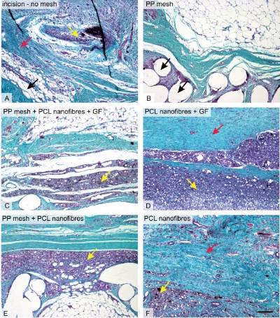
Histological evaluation. Collagen, adipose tissue, and granulomatous infiltration in the scaffolds under study. In samples without any mesh (A), the incision was healing with a mixture of collagen (black arrow), adipose connective tissue (red arrow) and inflammatory infiltrate (yellow arrow). Samples with polypropylene (PP) mesh (B) had a high fraction of adipose tissue, but the spaces showing the dissolved mesh (black arrows) were surrounded by only a few inflammatory cells. Remnants of the nanofibers (C, D, E, F) were surrounded by granulomatous leukocyte-rich connective tissue (yellow arrows in C, D, E, F). The highest fraction of collagen (red arrow) was in samples of PCL nanofibers with adhered growth factors (GF) (D), followed by samples with no mesh (A) and by samples of PCL nanofibers (F). Low fractions of adipose tissue were found in samples of PCL nanofibers with adhered GF (D), samples with no mesh (A) and in samples of PCL nanofibers (F).
|
Publication: Plencner M, East B, Tonar Z, Otáhal M, Prosecká E, Rampichová M, Krejčí T, Litvinec A, Buzgo M, Míčková A, Nečas A, Hoch J, Amler E, (2014): Abdominal closure reinforcement by using polypropylene mesh functionalized with poly-ε-caprolactone nanofibers and growth factors for prevention of incisional hernia formation. Int. J. Nanomed. 9: 3263-3277, IF 4.195
Important results in 2013
1.Time-regulated drug delivery system based on coaxially incorporated platelet alpha granules for biomedical use
Alpha granules are novel source of natural growth factors from platelets. In recent work we had sucesfully embedded alpha granules into nanofibers with core/shell structure. The alpha granules survived the electrospinning proces and growth factors retained their bioaktivity as was demonstarted on the model of chondrocytes and mesenchymal stem cells.
|
Fig.A.,B. Micrograph alpha granules encapsulated in coaxial nanofibers polycaprolactone and polyvinyl alcohol using scanning electron microscopy (FESEM).
|
Collaboration:
Ústav biofyziky, 2.lékařská fakulta Univerzita Karlova v Praze; Oddělení mechaniky, Fakulta aplikovaných věd, Západočeská univerzita v Plzni; Textilní fakulta, Katedra netkaných textilií, Technická univerzita v Liberci
Publication:
Buzgo M., Jakubova R., Mickova A., Rampichova M., Prosecka E., Kochova P., Lukas D., Amler E.: (2012) Time-regulated drug delivery system based on coaxially incorporated platelet alpha granules for biomedical use. Nanomedicine- UK. 8(7): 1137-1154. IF 5,26
2. A cell-free nanofiber composite scaffold regenerated osteochondral defects in miniature pigs
A novel drug delivery system was developed on the basis of the intake effect of liposomes encapsulated in PVA nanofibers. Time-controlled release of insulin and bFGF improved MSC viability in vitro. In addition, cell-free composite scaffolds containing PVA nanofibers enriched with liposomes, bFGF, and insulin were implanted into seven osteochondral defects of miniature pigs; control defects were left untreated. The cell-free composite scaffold enhanced migration of the cells into the defect, and their differentiation into chondrocytes; the scaffold was able to enhance the regeneration of osteochondral defects in minipigs.
|
Fig. Regeneration of osteochondral defect of miniature pig using a cell-free collagen type I/hyauronate sodium/fibrin gel containing polyvinyl alcohol nanofibers enriched with liposomes and growth factors (A) and untreated defect (B) 12 week after implantation. Alcian blue and PAS staining.
|
Collaboration:
Fakulta biomedicínského inženýrství, ČVUT v Praze, Ústav biofyziky, 2. LF UK v Praze, Fyziologický ústav AV ČR, v.v.i., Ústav stavebníctva a architektúry SAV, Textilní fakulta, Technická univerzita Liberec, Ústav živočišné fyziologie a genetiky AV ČR, v.v.i., Ústav histologie a embryologie, 2. LF UK v Praze, Student Science, s r.o.
Publication:
Filová E., Rampichová M., Litvinec A., Držík M., Míčková A., Buzgo M., Košťáková E., Martinová L., Usvald D., Prosecká E., Uhlík J., Motlík J., Vajner L., Amler E. A cell-free nanofiber composite scaffold regenerated osteochondral defects in miniature pigs. Int J Pharm. 2013 Apr 15;447(1-2):139-49. IF 3,458.
3. Electrospun core/shell nanofibers: a promising system for cartilage and tissue engineering
Alpha granules are novel source of natural growth factors from platelets. In recent work we had sucesfully embedded alpha granules into nanofibers with core/shell structure. The alpha granules survived the electrospinning proces and growth factors retained their bioaktivity as was demonstarted on the model of chondrocytes and mesenchymal stem cells.
|
Fig. Coaxial nanofibres of polyvinyl alcohol (core) and polycaprolactone (sheath) with incorporated labeled alpha-granules carboxy fl uorescein succinimidyl ester scanned by confocal microscopy.
|
Collaboration:
Ústav biofyziky, 2. LF UK v Praze; Univerzitní centrum energeticky efektivních budov, Buštehrad
Publication:
Amler E., Mickova A., Buzgo M. Electrospun core/shell nanofibers: a promising system for cartilage and tissue engineering? Nanomedicine (Lond). 2013 Apr;8(4):509-12. IF 5,26.


