Clinical applicatins
The ability to identify and characterize microorganisms (bacteria, eukaryotic cells) from minute sample volumes in a rapid and reliable way is the crucial first step in the classification of microbial infections. Ideal analytical techniques would require minimal sample preparation, permit automatic analysis of many serial samples, and allow rapid classification of these samples against a stable database. Current practice, however, is far from this ideal; a typical analytical procedure might require as long as a few days. With the lack of timely laboratory results, for example, inappropriate antibiotics might be prescribed which could be ineffective against the microorganisms responsible for the infection. Not only does the patient's condition deteriorate, but the use of inappropriate drugs may well contribute to the emerging problem of drug resistance in microorganisms.
It has been shown in several studies that Raman microspectroscopy is capable of rapid identification and discrimination of biological samples including medically relevant microorganisms (bacteria, yeast). This experimental technique employs a laser beam that is focused with a microscope objective in order to excite and collect Raman scattering spectra from a small volume of the sample. A typical Raman spectrum contains a wealth of information indicative of the cellular content of nucleic acids, proteins, carbohydrates, and lipids. Such a spectrum functions as a cellular ‘fingerprint’ and serves as a sensitive indicator of the physiological state of the cell. Raman spectra thus enable to differentiate cell types, actual physiological states, nutrient conditions, and phenotype changes. In principle, Raman spectroscopy requires measurement times on the order of minutes, and sample preparation can be short and extremely economical.
 Examples of Raman spectra of S. aureus (blue), S. epidermidis (orange) and their variants without carotenoids (S. aureus – yellow) and with carotenoids (S. epidermidis – violet). The spectra were vertically shifted in order to increase the visibility of details. Future Microbiology 12, 881-890, 2017 Examples of Raman spectra of S. aureus (blue), S. epidermidis (orange) and their variants without carotenoids (S. aureus – yellow) and with carotenoids (S. epidermidis – violet). The spectra were vertically shifted in order to increase the visibility of details. Future Microbiology 12, 881-890, 2017
We have been recently involved in the above mentioned microorganisms research and managed to published following papers dealing with different “real-world” applications of Raman spectroscopy where bacteria or yeast were in the focus of our investigations.
Pilát, Z.; Bernatová, S.; Ježek, J.; Kirchhoff, J.; Tannert, A.; Neugebauer, U.; Samek, O.; Zemánek, P. Microfluidic Cultivation and Laser Tweezers Raman Spectroscopy of E. coli under Antibiotic Stress. Sensors 2018, 18, 1623.
D. Prochazka, M. Mazura, O. Samek, K. Rebrosova, P. Porizka, J. Klus, P. Prochazkova, J. Novotny, K. Novotny, J. Kaiser: "Combination of laser-induced breakdown spectroscopy and Raman spectroscopy for multivariate classification of bacteria", Spectrochimica Acta Part B 139, 6-12, 2018
K. Rebrosovsa, M. Siler, O. Samek, F. Ruzicka, S. Bernatova, J. Jezek, P. Zemanek, V. Hola: "Differentiation between Staphylococcus aureus and Staphylococcus epidermidis strains using Raman spectroscopy", Future Microbiology 12, 881-890, 2017
K. Rebrosovsa, M. Siler, O. Samek, F. Ruzicka, S. Bernatova, V. Hola, J. Jezek, P. Zemanek, J. Sokolova, P. Petras:"Rapid identification of staphylococci by Raman spectroscopy", Scientific Reports 7, 14846, 2017
Mlynáriková, K.; Samek, O.; Bernatová, S.; Růžička, F.; Ježek, J.; Hároniková, A.; Šiler, M.; Zemánek, P.; Holá, V. Influence of Culture Media on Microbial Fingerprints Using Raman Spectroscopy. Sensors 2015, 15, 29635-29647.
Samek, O.; Mlynariková, K.; Bernatová, S.; Ježek, J.; Krzyžánek, V.; Šiler, M.; Zemánek, P.; Růžička, F.; Holá, V.; Mahelová, M. Candida parapsilosis Biofilm Identification by Raman Spectroscopy. Int. J. Mol. Sci. 2014, 15, 23924-23935.
Bernatová, S.; Samek, O.; Pilát, Z.; Šerý, M.; Ježek, J.; Jákl, P.; Šiler, M.; Krzyžánek, V.; Zemánek, P.; Holá, V.; Dvořáčková, M.; Růžička, F. Following the Mechanisms of Bacteriostatic versus Bactericidal Action Using Raman Spectroscopy. Molecules 2013, 18, 13188-13199.
|
Biotechnological application
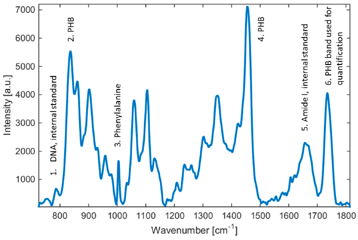
Recently, we have investigated an easy-to-apply Raman spectroscopy technique for the qualitative and quantitative determination of the amount of poly(3-hydroxybutyrate) (PHB) in bacteria Cupriavidus necator H16. The technique is applicable to near real-time and in-situ monitoring at the process control of PHB bio production. Here, Raman spectroscopy demonstrates clear benefits over existing chemical means of identification, such as gas chromatography in the speed and noninvasivity of the quantification process.

Raman spectra of Cupriavidus necator H16. Selected emission lines used in our study are highlighted. Sensors 2016, 16, 1808
Fast and reliable determination of intracellular PHB content during biotechnological production of PHB can take about 12 min. In contrast, gas chromatography analysis takes approximately 8 h.
Samek, O.; Obruča, S.; Šiler, M.; Sedláček, P.; Benešová, P.; Kučera, D.; Márova, I.; Ježek, J.; Bernatová, S.; Zemánek, P. Quantitative Raman Spectroscopy Analysis of Polyhydroxyalkanoates Produced by Cupriavidus necator H16. Sensors 2016, 16, 1808.
|


 Examples of Raman spectra of S. aureus (blue), S. epidermidis (orange) and their variants without carotenoids (S. aureus – yellow) and with carotenoids (S. epidermidis – violet). The spectra were vertically shifted in order to increase the visibility of details. Future Microbiology 12, 881-890, 2017
