Services
Routine Services
CHARACTERISTICS:
- readily available
- any compound is eligible for the screening
- carried out mostly by technical personnel according to standard operational procedures

RESPONSIBLE PERSONS:
Eva Tloušťová, ☎ 271 (cytotoxicity assays, sample receipt data handling and export)
Jana Günterová, ☎ 223 (flow cytometry)
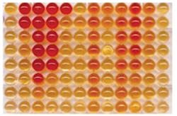
- Cytotoxicity screening: Routinely, we test the cytotoxicity of the compounds in four cancer cell lines of which two represent hematologic malignancies (CCRF-CEM, HL-60) and two are solid tumor-derived (HepG2, HeLa S3). We employ a well-established XTT colorimetric assay based on the measurement of the activity of mitochondrial dehydrogenases of respiring (i.e. living) cells. In case it is not possible to use this standard assay (e.g. colored, fluorescent or otherwise difficult samples) we also offer alternative analytical methods such as ATP luminescent assay or lactate dehydrogenase assay. If you need any other cell lines for your research, please contact us.
- Cell cycle analysis: Automatically, we perform cell cycle analysis in all compounds found to possess cytotoxic activity and we do that at IC50 value from the cytotoxicity assay (other concentrations upon request). The method is based on the flow cytometric analysis of DNA content following the staining of permeabilized cells with propidium iodide. Cell cycle arrest in its respective phases (G1/0, S, G2, M) is indicative for its mechanism of action. The assay helps to improve your papers focused on the anticancer activity of the compounds.
Special Services
CHARACTERISTICS:
- intended for limited series of compounds
- execution may take longer time depending on actual demand
- require input of qualified personnel

RESPONSIBLE PERSONS:
Helena Mertlíková Kaiserová, ☎ 114
Mirek Hájek, ☎ 393
Note: If you are using certain service repeatedly in the same experimental setup, you can address directly the person who physically performs this particular assay
- Apoptosis determination: Apoptosis is a process of programmed cell death. Apoptosis activation is particularly desirable in prospective anticancer drugs. It is often necessary to distinguish between apoptosis and necrosis, which is characterized by uncontrolled cell lysis and consequent leakage of its intracellular content into the environment. Our dual Annexin V / propidium iodide assay typically enables us to identify the mode of cell death induced by a compound. Quantification of the apoptotic/necrotic cell is flow-cytometry based. In addition, we offer alternative methods for apoptosis determination: caspase activation in situ with use of CaspACE fluorescent marker, caspase activity (3, 8, 9) in cell lysate, DNA fragmentation (DNA ladder), mitochondrial depolarization (FACS).
- RNA/DNA synthesis evaluation: To this end we employ a non-radioactive immunocytochemical method based on the incorporation of brominated nucleic acid precursors i.e. BrU and BrdU into the newly synthesized RNA/DNA strand. The cells are then stained with anti-BrU antibody and fluorescently labelled secondary antibody. The RNA/DNA synthesis is then quantified by flow cytometry.
- Detection of reactive oxygen species (ROS) and oxidative stress markers: We offer a simple flow cytometric method for quantification of intracellular ROS. The method is based on the oxidation (activation) of the fluorescein derivative CM-H2DCF-DA. The probe does not react with extracellular ROS in the reaction media since it is only functional following the activation by cellular esterases. Another probe we have in our portfolio, MitoSOX, is specifically designed for detection of mitochondrial superoxides. The presence of oxidative stress in the cell can also be quantified by determination of glutathione level or lipid peroxidation products (malondialdehyde – "TBARS assay").
- Intracellular Ca2+ determination: Measuring of free intracellular calcium level has numerous applications. Ca2+ plays an important role in cell signalling, neurotransmitter release from neuronal cells, contractions of all muscle cell types (e.g. cardiomyocytes) and it is also a target for various toxins. We employ Fura-2 fluorescent probe as Ca2+ sensor. The probe is only activated inside the cell where it emits green fluorescence following its binding to Ca2+. The signal is recorded and quantified using a plate reader.
- Mitochondrial membrane potential (Ψ) evaluation: Another simple flow cytometric method useful for determination of the mode of apoptotic cell death or mitochondrial toxicity assessment. To this end we use JC-1 carbocyanine dye, which accumulates in mitochondria and exists either as a monomer at low Ψ (green diffuse fluorescence) or forms J-aggregates at high Ψ (red punctate fluorescence).
- Caco-2 permeation test: The method is useful for prediction of intestinal permeability (e.g. bioavailability after oral administration) of the drug. The Caco-2 cells are derived from human colon carcinoma and are able to differentiate to form a polarized monolayer of the cells resembling the intestinal barrier (enzyme expression, tight junctions, brush border). The transport of the compound can be assessed in both directions (apical to basolateral side and vice versa) which serve as an indicator of active efflux. The quantification of the compounds following the assay is performed by HPLC (in some case MS).
-
Kinase assays: Kinases represent an important group of enzymes transferring the γ-phosphate from ATP to an acceptor molecule. In our laboratory we employ a universal and sensitive luminescent kit based on the detection of ADP formed. Theoretically, any pair of substrate/kinase can be detected. At the moment we offer screening for inhibitors of two lipid kinases (PI4KA, PI4KB). Other kinase assays may be developed depending on the actual needs (limiting factor: availability and price of the enzyme).
Note: The service is not meant for broad kinase profiling of the compounds. This has to be tested commercially. - Inhibition of various enzymes of purine/pyrimidine metabolic pathways: Biochemistry of modified nucleos(t)ides and nucleobases has been in the focus of the group for a long time. We have a number of functional and sensitive methods at our disposal (mostly radiometric) for detection of the activity/inhibition of xanthine oxidase, thymidine phosphorylase, adenosine deaminase, adenosine kinase, SAH-hydrolase, purine nucleoside phosphorylase and others. (limiting factor : availability and price of the enzyme and labelled substrate).
- NS5B polymerase inhibition: Key enzyme of HCV replication. Enzyme activity is determined by means of classical polymerase reaction with use of specific RNA template and the mix of nucleotides. Detection is based on quantification of incorporated α-33P-GTP on phosphorimager. As this is not a cell-based assay, it is critical to provide the inhibitors in their active form i.e. triphosphate analogs. The results do not necessarily correlate with anti-HCV activity of the compounds.
- Isolation and cultivation of primary cells: Classical immortalized cell lines may not be suitable for some specific applications where terminally differentiated cells are desired. Currently, we have developed techniques for isolation of mouse embryonal neuroblasts which are then differentiated into the mature neuronal/glial cells. These cultures are used for studying the neuroprotective compounds. Also, we have expertise in the isolation and culture of other primary cell types (cardiomyocytes, hepatocytes). Limiting factor: availability of the animals.
- Mitochondrial toxicity assessment: Recommended for the compounds that proved to be biologically active towards their intended targets and seem to be promising for the next phase development as prospective drugs. Early detection of mitochondrial toxicity may prevent late-stage drug attrition. Standard cytotoxicity testing is not sufficient for identification of mitotoxic compounds!! We offer mitochondrial toxicity assessment by Glu/Gal assay, which is based on comparison of compound toxicity in glucose- vs. galactose-containing culture medium. The absence of glucose shifts cellular metabolism from glycolysis towards oxidative phosphorylation and subsequent sensitization to the mitochondrial toxicants. Determination of the effects of the compounds on mtDNA content and/or mtDNA-encoded protein expression is particularly useful for nucleos(t)ide analogs. The assay is currently in preparation.
- Dual luciferase reporter assay: The assay is commonly used as a tool to study gene expression at the transcriptional level. Two reporter genes (experimental and control) are transfected in the appropriate cell line while experimental reporter expression is controlled by the regulatory region of interest. Two distinct luciferases (firefly and Renilla l.) are used as reporters. The use of control reporter is necessary for data normalization thus eliminating non-relevant influences such as compound cytotoxicity.