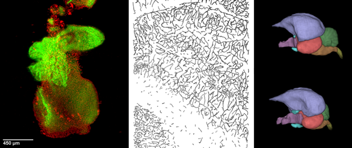
The Department of Biomathematics consists of two research groups: (1) Bioimaging and Image Analysis, and (2) Biochemistry of Membrane Receptors. Operating mainly at in situ and in vitro levels, respectively, the two groups complement each other when studying phenomena confined to the cell membrane.
1. Bioimaging and Image Analysis
The group conducts research into the 3D microanatomical aspects of physiological phenomena at mesoscopic, microscopic and ultrastructural level. It is engaged in a range of collaborative projects across the campus and beyond. If interested please contact us (jiri.janacek@fgu.cas.cz).
CURRENTLY FUNDED PROJECTS
- Brain development in birds, as relevant to the ability to fly
- The conduction system of developing murine heart
LONG-TERM PROJECTS
- Development of 3D image analysis and stereological methods including software (employed, e.g., in a volumetric analysis of Langerhans islets from human donors)
- Microscopic optical-contrasting (label-free) strategies to visualize living cells and tissues
OTHER PROJECTS (selection only)
- Effects of ionizing radiation on blood capillaries network
- Effects of elevated CO2 on the photosynthetic apparatus’ performance and ultrastructure
- Mechanisms governing the formation of extracellular matrix proteins (collagen and elastin)
IN-HOUSE IMAGING MODALITIES
- Laser scanning confocal microscopy
- Two-photon excitation microscopy
- Second harmonic generation (SHG) microscopy
- ‘White-light‘ laser and hybrid detectors (on Leica TCS SP8 WLL MP microscope)
- FRAP recording, FRET analysis, FLIM/PLIM recording including λ2 scans
- Optical projection tomography (OPT)
- Selective plane illumination (light sheet) microscopy (SPIM)
- Phase-, modulation- and interference contrast microscopy
AVAILABLE SOFTWARE AND PROTOCOLS
- Software for 3D image analysis, stereology, spatial statistics and deconvolution (Hyughens)
- Optical/chemical clearing protocols for OPT and SPIM
OPEN ACCESS TO EQUIPMENT
The group is part of the Czech BioImaging infrastructure [1] supporting open access to most of its microscopes (listed above), and implicitly also to its expertise. The portfolio includes, among other imaging equipment located at other departments, a Nipkow spinning-disk (“Carv II“) confocal microscope [2].
[1] http://www.czech-bioimaging.cz/
[2] http://www.fgu.cas.cz/en/articles/529-czech-bioimaging-2016-2019
2. Biochemistry of Membrane Receptors
Our research focuses on the cellular and molecular mechanisms of desensitization of hormone response mediated by the activation of G protein coupled receptors (GPCR). G protein coupled receptors are plasma membrane integral proteins which serve as transducers of extracellular signals across the plasma membrane bilayer to the cell interior. GPCR play a key role in the regulation of many physiological processes and functions. Moreover, GPCRs represent one of the most important groups of targets for therapeutics.
The main areas of the research are:
- The role of cell membrane and membrane domains
- Opioid receptors and drug addiction
- The effect of monovalent ions on δ-opioid receptors – analysis of lithium effect in living cells and isolated cell membranes
- GABA-B - signalling cascades


