

Laboratoř materiálových analýz a mikroskopie
Ústav teoretické a aplikované mechaniky › CET › O CET › Laboratoře › Laboratoř materiálových analýz a mikroskopie
Lidé:
Vedoucí laboratoře:
Ing. Alberto Viani, Ph.D.
viani@itam.cas.cz
+420 567 225 308
Členové laboratoře:
Mgr. Petra Mácová
Eva Pažourková
Ing. Konstantinos Sotiriadis, Ph.D.
Ing. Milan Svoboda
Mgr. Radek Ševčík, Ph.D.
Bruker D8 Advance diffractometer (Bragg-Brentano theta-theta geometry)
Specifications
- Ni-filter
- LYNXEYE Position Sensitive Detector
- rotating sample stage
- multi-purpose sample stage for massive samples
- Euler cradle for microstructural measurements
- parallel beam optics
Applications
This is the technique of election for investigating solid samples. It’s mainly used with samples in form of powders. Sample preparation can be accomplished with our crushing/milling equipment. Typical use is execution of qualitative analysis: phase identification in polycrystalline samples, identification of minerals in geological samples, detection of polymorphs in pharmaceutical samples, the determination of impurities in a pure phase (down to 0.1 weight %).
More advanced data treatment (Rietveld method) allows also to perform powder diffraction quantitative analysis: Quantification of phases into powdered samples, like minerals in geological samples, in raw materials, in industrial ceramics, refractories, cements, mortars. The technique is suitable for the quantification of non-crystalline component (amorphous) as well.
X-ray Crystallography: investigation of the crystal structure of known and unknown phases.
Crystallite size and residual strain: the determination of mean dimension of diffraction domains (crystallites) allows for the characterisation of samples undergoing thermal or mechanical treatments.
Texture analysis: the reconstruction of the distribution of orientation of the crystallites in polycrystalline samples, allows for the reconstruction of the sample microstructure. Specific orientation of crystallites develops as a result of industrial production processes (as for many metallic parts) or by action of external or internal agents. Examples are the way in which carbonate crystals grow in molluscs shells, the way in which crystal habit develops in rocks subjected to oriented pressure (metamorphism), the way in which crystals grow within processed technical ceramic bodies or metals.
Grazing incidence: with this technique the identification of phases deposited at the surface of solid samples in form of films in the range 10-200nm, and their thickness, can be investigated. Typical field of application are materials for electronics, functional materials.
Peculiar to the last two methods is that they are non destructive, the sample is analysed without preparation with the aid of specific sample stages.
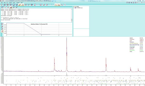
Typical output of Rietveld refinement of X-ray powder diffraction data. On the lower half of the picture, the original spectrum (in blue) and the refined one (in red) together with the difference between the experimental and the calculated (gray line) and markers corresponding to reflection peaks of the phases in the sample, are depicted. Quantification of all the phases including the amorphous (non crystalline) fraction is reported on the bottom right.
SEM FEI Quanta 450 FEG (scanning electron microscope)
Specifications
- high resolution Schottky field emission emitter
- accelerating voltage: 200 V to 30 kV
- magnification: 6 to 1000000x
Detectors
- Everhardt Thornley SED (secondary electron detector)
Backscattered electron detector
- Large Field Low vacuum SED (LFD)
- Gaseous SED (GSED) (used in ESEM mode)
- IR camera for viewing sample inside the chamber
- EDS spectrometer
Cathodoluminescence detector
- EBSD detector
Main features
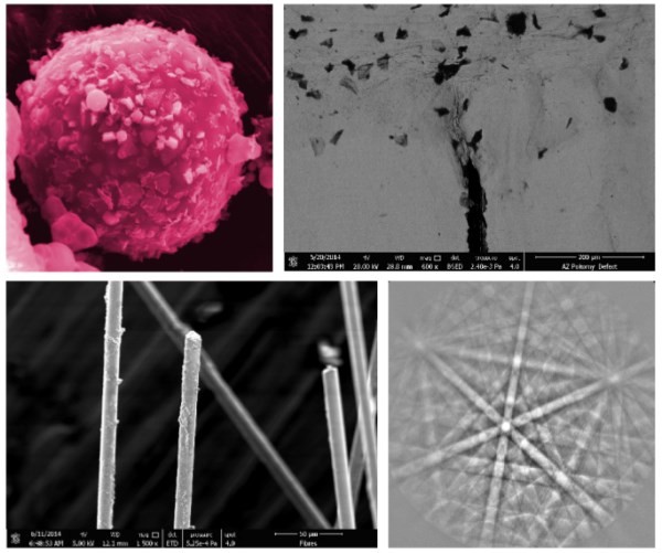
The microscope is equipped with the last generation emitter, allowing for a stable flux of electrons and high intensity. This is a versatile instrument that can be used to investigate a wide range of specimens ranging from inorganic to organic. Although the best results are obtained with a superficial conductive coating of few nanometers (usually gold or carbon), it’s possible to characterize even non-conductive samples, avoiding any treatment.
Working under low vacuum and ESEM enables charge-free imaging and analysis of non-conductive and/or hydrated specimens.
Applications
NanoCharacterization: Metals & alloys, oxidation/corrosion, fractures, failure analysis, welds, polished sections, magnetic and superconducting materials. e.g. ceramics, composites, plastics, films/coatings, geological sections, minerals, soft materials (polymers, pharmaceuticals, filters, gels, tissues, plant material), particles, porous materials, cements, fibers.
In situ NanoProcesses: hydration/dehydration; wetting behaviour/contact angle analysis; oxidation/corrosion; tensile (with heat or cooling); crystallization/phase transformation.
The EBSD detector, allows for accomplishing micro-diffraction on the specimen’s surface, thus, identify the nature of crystalline phases within the sample and perform texture analysis: the reconstruction of the distribution of orientation of the crystallites in polycrystalline samples, allows for the reconstruction of the sample microstructure. Specific orientation of crystallites develops as a result of industrial production processes (as for many metallic parts) or by action of external or internal agents. Examples are the way in which carbonate crystals grow in molluscs shells, the way in which crystal habit develops in rocks subjected to oriented pressure (metamorphism), the way in which crystals grow within processed technical ceramic bodies or metals. The EDS detector allows for investigating the chemical composition with high spatial resolution.
- autosampler with 49 positions
- anions – fluoride, chloride, bromide, nitrite, nitrate, phosphate, sulphate
- cations – lithium, sodium, ammonium, potassium, magnesium, calcium

- room temperature DTGS and liquid nitrogen-cooled MCT detectors
- transmission, ATR and DRIFT techniques
- ATR with diamond crystal inside
- DTGS detecto
Typical applications include: identification of compounds following their thermal decomposition/transformation with temperature (e.g. phases of hydration in mortars nad concrete), and their quantification; determination of decomposition temperatures, glass transition temperature in polymers, calculation of specific heat capacity. Engine oil volatility measurements, filler content, flammability studies, measurement of volatiles (e.g. water, oil), oxidative stabilities, thermal stabilities, catalyst and coking studies, melting/crystallization behavior. The technique is particularly suitable for the analysis of construction, ceramic and geological materials.
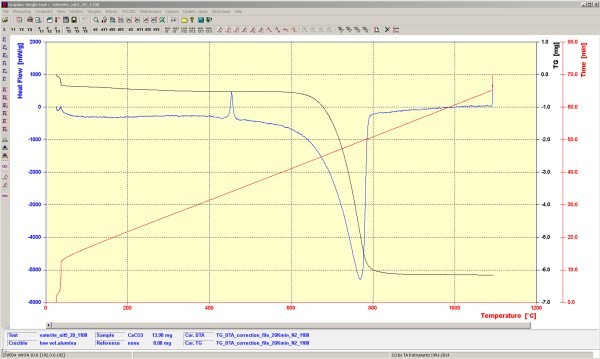
In this image, corresponding to a sample of CaCO3, a phase transition is shown as an exothermic peak (up) at about 450°C and the decomposition as an endothermic peak (down) with a weight loss at 750°C. These results allows to determine and quantify the composition of the sample.
- stanovení porozity pevných materiálů
- rtuť je pod kontrolovaným tlakem vtlačována do pórů materiálu a podle rychlosti a potřebného tlaku jsou zjišťovány charakteristiky vzorku
- přístroj poskytuje informace o distribuci velikosti pórů, analýze tvaru pórů, poměr velikosti ústí a objemu pórů, permeabilitě a stlačitelnosti materiálu, turtuositě pórů, fraktálních rozměrech atd.
- měření v rozsahu tlaků od 0 do 228 MP
- rozsah: průměr pórů 5nm – 360 µm
- stanovení měrné hmotnosti
- vzorek je vložen do komory o známém objemu, hélium o určitém tlaku je vpuštěno do referenční komory a vzápětí do měřící komory, kde se souběžně měří jeho tlak, ze vzájemných poměrů tlaků a objemů se spočítá hustota matrice materiálu
- velikost vzorku 0,01-350cm3
- stanovení měrného povrchu a distribuce velikosti pórů pevných materiálů
- přístroj využívá principu fyzikální adsorpce k získání adsorbčních a desorpčních izoterem a k získání informacích o povaze zkoumaného materiálu
- dva plně automatizované odplyňovací systémy s řízenou dobou zahřívání
- dvanáct plynových přívodů, které jsou automaticky nastavitelné
- software s velkou flexibilitou naměřených dat
- slouží k dokončování obrábění, díky tomuto přístroji je možné dosáhnout vysoké přesnosti rozměrů i geometrického tvaru a povrchů s nejnižší drsností
- plně programovatelná bruska/leštička
- dotykový panel na řídící konzole, možnost využití naprogramovaných postupů a systému Zaxis (umožňuje nahradit dobu pro krok mírou úběru materiálu)
- elektro-hydraulický systém, který umožňuje definovat tlak, teplotu topení a dobu chlazení
- formy pro lisování o velikostech od 25 mm do 50 mm
- krátký čas pro cyklus přípravy vzorku
- zařízení pro současnou infiltraci a zalévání většího počtu vzorků
- obsahuje vakuovou pumpu se stabilním zásobníkem vakua, zabudovaný synchronní motor, licí zařízení, manometr, filtr pro pohlcování vlhkosti
- popis složek materiálu, velikosti, tvaru, distribuce jednotlivých složek, mineralogického složení
- digitální mikroskop pro pozorování s velkou hloubkou ostrosti při umístění na stativu nebo při držení v ruce
- morfologie neupravených povrchů
- petrografické rozbory v průchozím polarizovaném světle
- mikroskop určený pro materiálové vědy se zvětšením až 120x