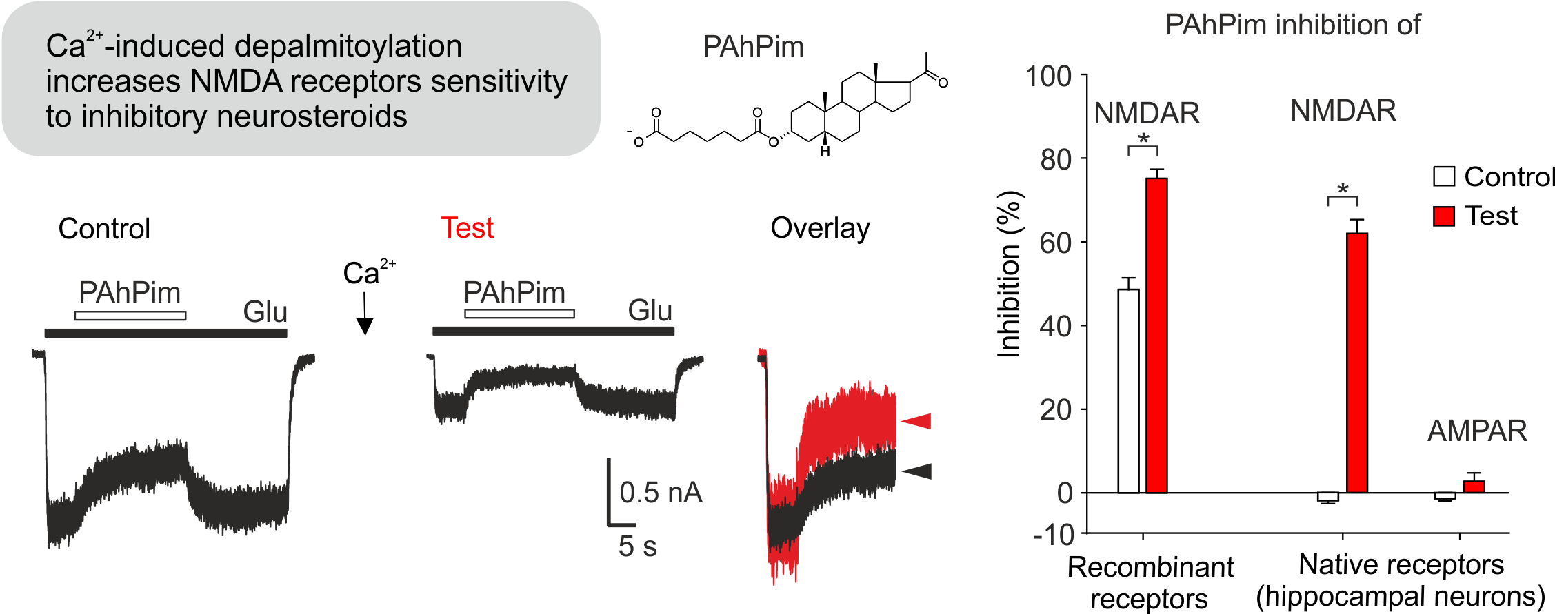
Palmitoylation controls NMDA receptor susceptibility to neurosteroids (3.5. 2021)
N-methyl-D-aspartate (NMDA) receptors are ionotropic glutamate receptors that are crucial for synaptic transmission, learning, and memory acquisition. Their overactivation leads to pathology associated, for example, with stroke or Alzheimer's disease. Overactivation of NMDA receptors can be inhibited by a number of substances, including neurosteroids.
Using electrophysiological and molecular-biological techniques, we have elucidated the molecular mechanism by which NMDA receptor susceptibility to inhibitory neurosteroids is increased. This change is due to depalmitoylation of three cysteines (C849, C854, C871) in the intracellular part of the GluN2B receptor subunit, which occurs after a transient increase in intracellular concentration of Ca2+. Beyond the pharmacological consequences, depalmitoylation of the receptor results in a change in kinetic parameters in favor of the closed state. Increased sensitivity of NMDA receptors to inhibitory neurosteroids is thus another of the neuroprotective mechanisms that prevents excitotoxic damage to nerve tissue.
Hubálková, Pavla - Ladislav, Marek - Vyklický, Vojtěch - Smejkalová, Tereza - Hrčka Krausová, Barbora - Kysilov, Bohdan - Krůšek, Jan - Naimová, Žaneta - Kořínek, Miloslav - Chodounská, Hana -Kudová, Eva - Černý, Jiří - Vyklický ml., Ladislav Palmitoylation Controls NMDA Receptor Function and Steroid Sensitivity. Journal of Neuroscience 2021. 41 (10) 2119-2134, F: 5.674 DOI
Clove oil alleviates cold-induced toothache by blocking the TRPC5 ion channel (30.4. 2021)
Touching a cold drink can be a suffering for us when we have tooth decay. An international team of scientists led by Prof. Katharina Zimmermann (Friedrich - Alexander University Erlangen - Nuremberg in Germany), together with scientists from the Institute of Physiology of the Czech Academy of Sciences in Prague, found out how teeth detect cold and determined the molecular basis of cold-induced dental pain. In both mice and humans, dental cells called odontoblasts contain special cold sensors, the TRPC5 ion channels, which transmit information about a painful stimulus to the nervous system. The study also offers an explanation for why clove oil, which has been used for centuries in dentistry, can alleviate toothache. Clove oil contains a chemical eugenol that blocks the TRPC5 protein and prevents it from activating nerves. Video

Odontoblasts containing the ion channel TRPC5 (green) tightly pack the area between the pulp and the dentin in a mouse’s molar. The cells’ long-haired extensions fill the thin canals in dentin that extend towards the enamel. Sensory nerves are indicated in red (bIII-tubulin). Cell nuclei are stained with Hoechst 33258. (Credit: L. Bernal et al./Science Advances 2021)
Bernal, L. - Sotelo-Hitschfeld, P. - König, Ch. - Sinica, Viktor - Wyatt, A. - Winter, Z. - Hein, A. - Touška, Filip - Reinhardt, S. - Tragl, A. - Kusuda, R. - Wartenberg, P. - Sclaroff, A. - Pfeifer, J. D. -Ectors, F. - Dahl, A. - Freichel, M. - Vlachová, Viktorie - Brauchi, S. - Roza, C. - Boehm, U. - Clapham, D. E. - Lennerz, J. K. - Zimmermann, K. Odontoblast TRPC5 channels signal cold pain in teeth. Science Advances. Roč. 7, č. 13 (2021), č. článku eabf5567. ISSN 2375-2548. IF: 13.117, rok: 2019. DOI
Nanocellulose as a promising cell carrier for applications in regenerative medicine (8.4. 2021)
Cellulose in the form of a fabric has been used for thousands of years as a traditional wound dressing material. Nowadays, it can be used not only for passive wound covering, but also for active wound healing, e.g. with controlled delivery of various drugs or cells for regenerating the damaged tissue. A suitable form of cellulose for this purpose is nanocellulose, i.e. cellulose in the form of nanofibrils, simulating the architecture of the native extracellular matrix. This form of cellulose is produced by some species of bacteria, or can be isolated from higher plants, including wood. In our experiments, we have focused on the development of “intelligent” wound dressings, capable of delivering skin and stem cells into skin wounds. These dressings are based on electrically-charged cellulose nanofibrils, attached to a microfibrous cellulose fabric. Anionic nanocellulose provided a suitable substrate for the adhesion and growth of human dermal fibroblasts and human adipose tissue-derived stem cells, while cationic nanocellulose provided better support for cell-cell adhesion and for the formation of cell aggregates, which was apparent mainly in fibroblasts (Fig. 1). This difference was due to the preferential adsorption of albumin from the serum supplement of the culture medium, which is non-adhesive for cells, on cationic nanocellulose. However, both types of nanocellulose are useful in regenerative medicine. Anionic nanocellulose is suitable for creating continuous cell sheets, which can be delivered into the wound either spontaneously or after release from the substrate using cellulase enzymes, while cationic cellulose is suitable for creating cell spheroids, i.e. important structures for developing organoids and for tissue engineering.

Fig. 1 Morphology of normal human dermal fibroblasts (NHDFs; A) and human adipose tissue-derived stem cells (ADSCs; B), guided by the topography of cellulose meshes coated with anionic or cationic cellulose nanofibrils (a600, c600, respectively) after seven days of cultivation. 3D projection of microscopy images (front view and side view) of the cells on the material. F-actin of the cell cytoskeleton is stained in red, vinculin in the cells is stained in green. Confocal microscope with objective magnification 40x.
Pajorova J, Skogberg A, Hadraba D, Broz A, Travnickova M, Zikmundova M, Honkanen M, Hannula M, Lahtinen P, Tomkova M, Bacakova L, Kallio P. A cellulose mesh with charged nanocellulose coatings as a promising carrier of skin and stem cells for regenerative applications. Biomacromolecules, 2020, 21: 4857-4870, https://dx.doi.org/10.1021/acs.biomac.0c01097; IF = 6.092; DOI
Fat mass is controlled by the balance of triacylglycerol (TAG) degradation and synthesis. Adipose triglyceride lipase (ATGL) and hormone-sensitive lipase (HSL) are key players in TAG catabolism providing fatty acids (FAs) as energy substrates and metabolic intermediates. In collaboration with colleagues from University of Graz, Université de Montpellier and Institute of Organic Chemistry and Biochemistry, Czech Academy of Sciences, we discovered that ATGL and HSL metabolize TAGs containing antidiabetic lipid mediators (FA esters of hydroxy FAs), distinctly controlling the release of bioactive lipids. Our paper connects lipolysis-mediated TAG metabolism with the regulation of antidiabetic signaling lipids.
Glycerol-bound FAHFAs in TAG, named TAG estolides, serve as metabolite reservoir of FAHFAs mobilized by lipases upon demand. We found that ATGL alone or co-activated by comparative gene identification-58 (CGI-58) efficiently liberated FAHFAs from TAG estolides with a preference for more compact substrates where the estolide branching point is located near the glycerol ester bond. ATGL was further involved in transesterification and remodeling reactions leading to the formation of TAG estolides with alternative acyl compositions. HSL represented a much more potent estolide bond hydrolase for both TAG estolides and free FAHFAs. FAHFA and TAG estolide accumulation in white adipose tissue of mice lacking HSL argued for a functional role of HSL in estolide catabolism in vivo.
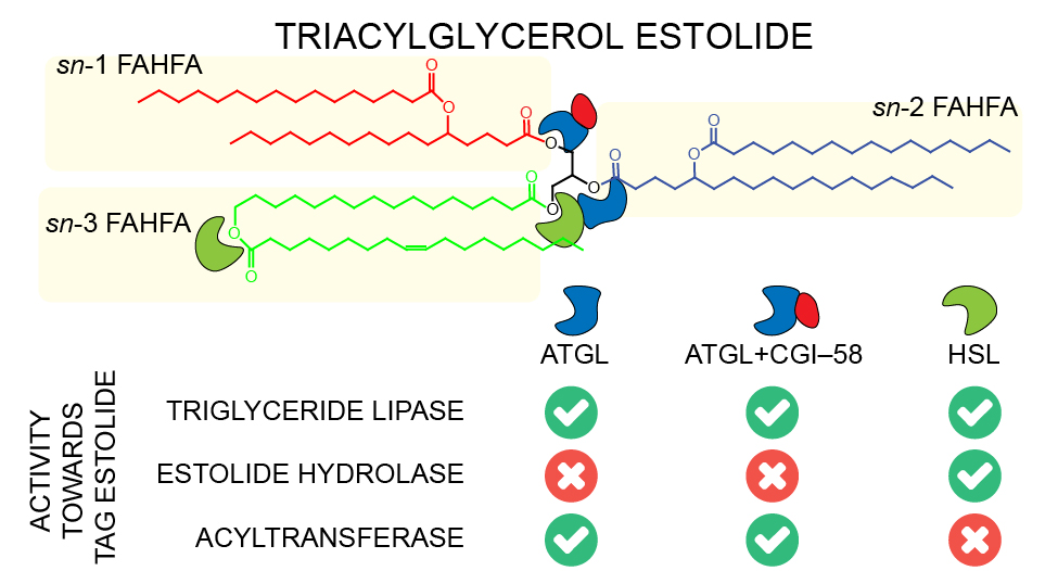
K. Brejchova, F.P.W. Radner, L. Balas, V. Paluchova, T. Cajka, H. Chodounska, E. Kudova, M. Schratter, R. Schreiber, T. Durand, R. Zechner, O. Kuda. Distinct roles of adipose triglyceride lipase and hormone-sensitive lipase in the catabolism of triacylglycerol estolides. Proceedings of the National Academy of Sciences of the United States of America 118(2) (2021) e2020999118. IF = 9.412 DOI
Mitochondrial cardiolipin in adipose tissue as a novel target for treatment of cardiac cachexia (7.12. 2020)
More than a half of deaths in the Czech Republic is due to the cardiovascular disease, with most of the patients suffering from heart failure (HF). Long‐term prognosis of the HF-patients worsens by cachexia, which develops in a subgroup of the patients. Effective treatment of cachexia is missing. In collaboration of scientists at the Institute of Clinical and Experimental Medicine and the Institute of Physiology of the Czech Academy of Sciences in Prague, a study was conducted in advanced HF-patients (n = 52) undergoing heart transplantation. In adipose tissue from the vicinity of the diseased hearts of the cachectic patients, we have demonstrated (i) stronger neurohumoral stimulation of fatty acid (FA) release, and (ii) synthesis of unusual phospholipid cardiolipin (CL 70:6). This phospholipid may deteriorate mitochondrial functions in fat cells and accelerate wasting of the tissue. It could serve as a novel therapeutic target in cachexia.
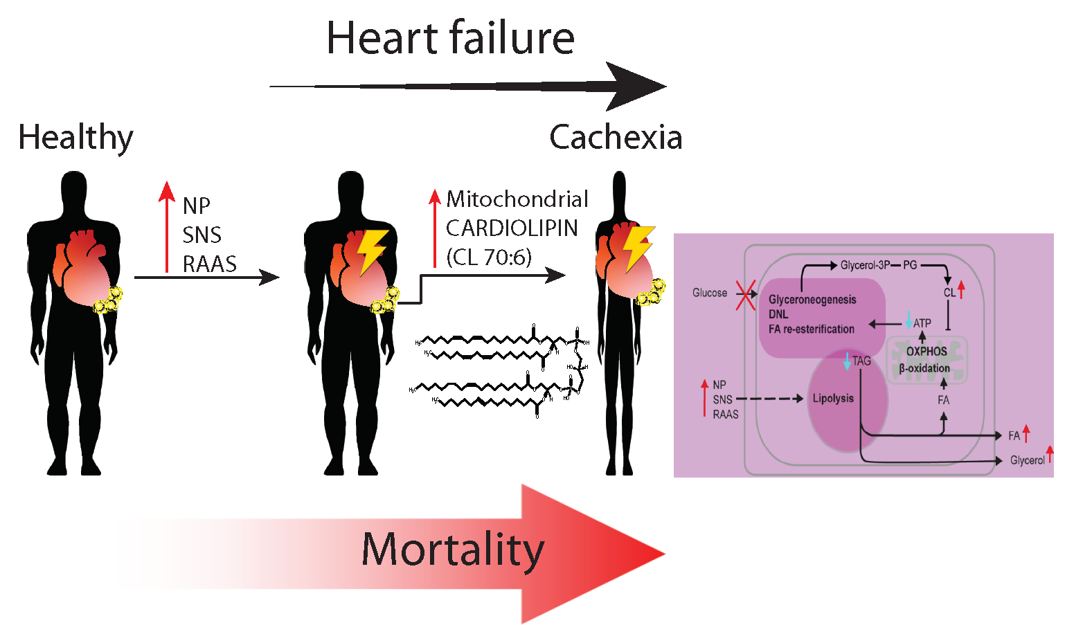
In HF patients, lipolytic breakdown of triacylglycerols (TAG) in adipose tissue is activated in response to natriuretic peptides (NP), sympathetic nervous system (SNS) and renin-angiotensin-aldosteron system (RAAS). This results in increased release of FA and glycerol into circulation. In body weight-stable patients, the breakdown is balanced by TAG synthesis, which depends on glyceroneogenesis, de novo synthesis of FA (DNL), and FA re-esterification. These reactions require ATP, which is synthesized by oxidative phosphorylation (OXPHOS) linked to β-oxidation of FA in mitochondria. In the cachectic patients, lipolysis is overstimulated, in association with synthesis of CL 70:6 in adipose tissue. This lipid induces uncoupling of OXPHOS and thus inhibits synthesis of ATP, resulting in insufficient replenishment of the TAG pool in fat cells and wasting of adipose tissue.
Janovska P, Melenovsky V, Svobodova M, Havlenova T, Kratochvilova H, Haluzik M, Hoskova E, Pelikanova T, Kautzner J, Monzo L, Jurcova I, Adamcova K, Lenkova L, Buresova J, Rossmeisl M, Kuda O, Cajka T, Kopecky J. Dysregulation of epicardial adipose tissue in cachexia due to heart failure: the role of natriuretic peptides and cardiolipin. Journal of Cachexia, Sarcopenia and Muscle 2020; IF: 9.802 DOI
Neurosteroid pregnenolone sulfate - revealing of its site of action at the N-methyl-D-aspartate receptor (26.10. 2020)
NMDA receptors are ion channels involved in the transmission of electrical signals between nerve cells. Their insufficient function contributes to severe neuropsychiatric disorders such as schizophrenia or autism. NMDA receptor activity can be increased by a variety of compounds, including neurosteroids. In collaboration with the Institute of Organic Chemistry and Biochemistry CAS, we identified the binding site and elucidated the mechanism of action of the naturally occurring neurosteroid pregnenolone sulfate (PES). PES binds to the interface of membrane domains of the NMDA receptor in the closed state, and subsequent activation of the receptor by glutamate leads to a rearrangement of the membrane domains and stabilization of the open state of the NMDA receptor ion channel. These findings may contribute to the development of new drugs for the treatment of diseases associated with the dysfunction of the glutamatergic system.
The interaction of the PES molecule (green) with the membrane domains of the NMDA receptor in the open state was obtained by docking followed by molecular dynamics simulation. Amino acids involved in the interaction with the PES molecule are highlighted in red.

Hrčka Krausová B, Kysilov B, Černý J, Vyklický V, Smejkalová T, Ladislav M, Balík A, Kořínek M, Chodounská H, Kudová E, Vyklický ml. L. Site of Action of Brain Neurosteroid Pregnenolone Sulfate at the N-Methyl-D-Aspartate Receptor. Journal of Neuroscience. 2020; 40 (31) 5922-5936. IF: 5.673 DOI
Dietary supplementation with Krill oil as a novel strategy to improve insulin sensitivity and reduce liver fat accumulation in obesity (20.10. 2020)
Lifestyle interventions including healthy nutrition represent an essential part of prevention and treatment strategies for metabolic sequelae of obesity. Krill oil as an extract from the Antarctic krill Euphausia superba (i.e. crustaceans found in Antarctic waters) is a relatively new source of omega-3 polyunsaturated fatty acids (Omega-3). It has been shown that dietary supplementation with Omega-3, usually as part of a triacylglycerol- or ethyl ester-based concentrate, may reduce inflammation and excessive amounts of fat in the liver (i.e. hepatic steatosis).
However, in obese type 2 diabetic patients, the use of Omega-3 may not necessarily improve insulin sensitivity and may impair long-term control of glucose metabolism. Therefore, we tested in obese mice whether supplementation with Omega-3 using an alternative lipid carrier, i.e. phospholipids from Kril oil, could have beneficial metabolic effects while also improving glucose metabolism and insulin sensitivity.
Our studies showed that Krill oil supplementation was able to reduce hepatic steatosis to a greater extent than Omega-3 given as triacylglycerols, and this effect of Krill oil was associated with improved insulin sensitivity in the liver and at the whole-body level. The beneficial effect of Krill oil on glucose homeostasis was associated not only with the improved bioavailability of Omega-3 (e.g. EPA, n-3 DPA) in tissues, but also with circulating levels of palmitoleic acid, a previously identified lipokine (i.e. hormone of a lipid nature) with insulin-sensitising effects whose content in Krill oil is incerased. In addition, Krill oil more effectively then Omega-3 triacylglycerols induced catabolism of fatty acids from the diet directly in the intestine, which could contribute to its strong antisteatotic effects in the liver. Our findings provide a general rationale for using Omega-3-containing phospholipids as nutritional supplements with potent insulin-sensitizing and antisteatotic effects.
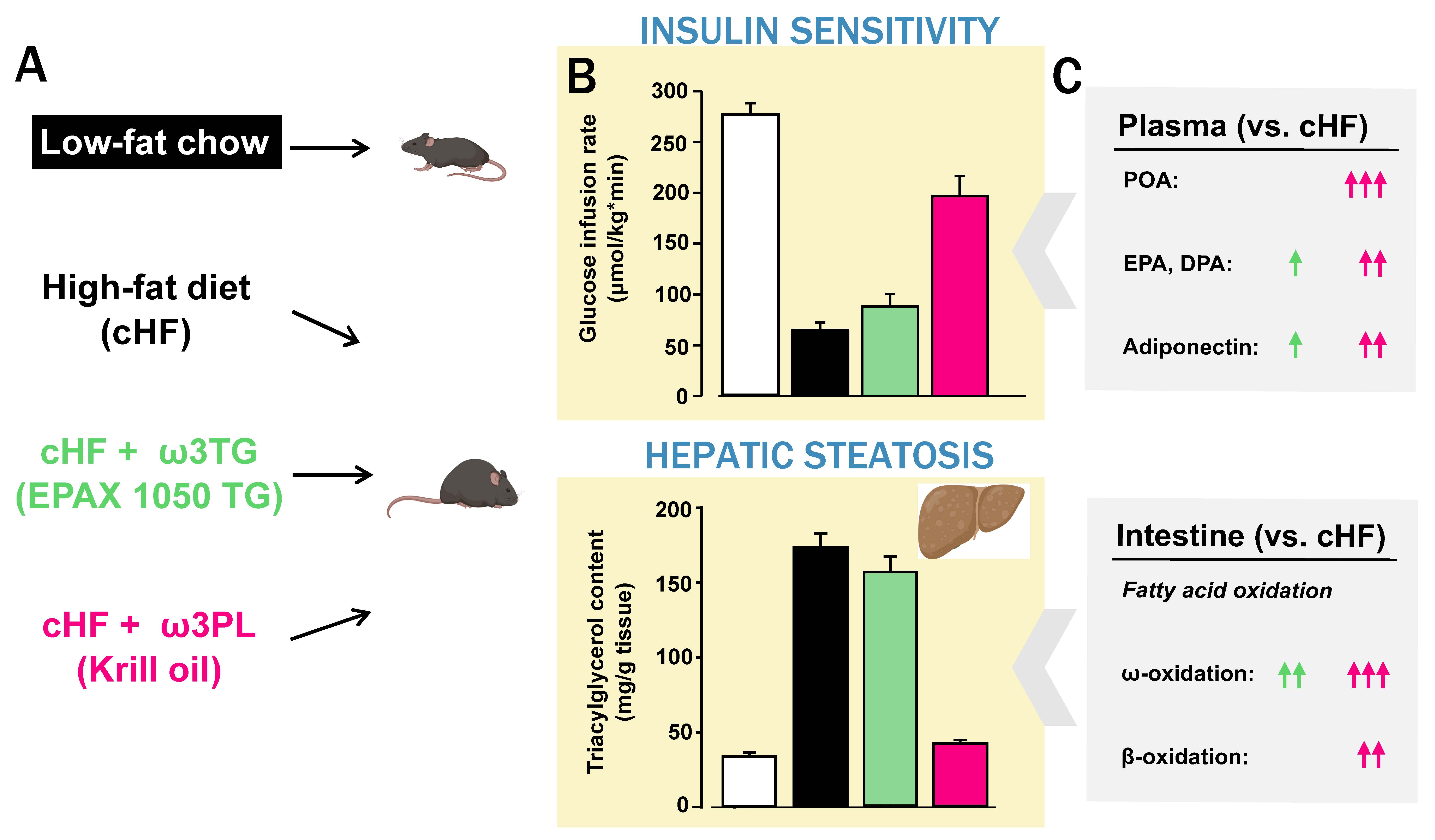
Male C57BL/6N mice were fed for 8 weeks a corn oil-based high-fat diet (cHF) alone or supplemented with Omega-3 contained in triacylglycerols (cHF+ω3TG) or Krill oil phospholipids (cHF+ω3PL); lean controls were fed a low-fat standard chow (A). Administration of cHF worsened insulin sensitivity (determined by measuring the glucose infusion rate during the hyperinsulinemic-euglycemic clamp; B – upper part) and caused significant accumulation of lipids in the liver (i.e. hepatic steatosis; B – lower part). While insulin sensitivity was almost preserved and hepatic steatosis virtually eliminated in cHF+ω3PL mice, much less pronounced effects were observed in cHF+ω3TG mice. Favorable metabolic changes observed after Krill oil administration may be related to increased circulating levels of insulin-sensitizing lipokine palmitoleate (POA; C – upper part) and increased fatty acid catabolism directly in the intestinal tissue (POA; C – lower part).
Rossmeisl M, Pavlisova J, Bardova K, Kalendova V, Buresova J, Kuda O, Kroupova P, Stankova B, Tvrzicka E, Fiserova E, Horakova O, Kopecky J. Increased plasma levels of palmitoleic acid may contribute to beneficial effects of Krill oil on glucose homeostasis in dietary obese mice. Biochim Biophys Acta Mol Cell Biol Lipids. 2020;1865(8):158732. IF: 4.519 DOI
Kroupova P, van Schothorst EM, Keijer J, Bunschoten A, Vodicka M, Irodenko I, Oseeva M, Zacek P, Kopecky J, Rossmeisl M, Horakova O. Omega-3 Phospholipids from Krill Oil Enhance Intestinal Fatty Acid Oxidation More Effectively than Omega-3 Triacylglycerols in High-Fat Diet-Fed Obese Mice. Nutrients. 2020;12(7):2037. IF: 4.546 DOI
Novel mechanism of insulin secretion highlights the importance of redox signalization (14.10. 2020)
Insulin secretion by β cells of pancreatic islets is essential for the maintenance of body glucose homeostasis. Its disturbance leads to the development of diabetes. Till now the mechanism of secretion has been linked exclusively to increased concentration of cellular energy molecule ATP initiating the cascade of insulin secretion. Our results showed that besides ATP the insulin secretion requires redox signalization. Specifically, we point out NADPH oxidase, isoform 4, which activity increases upon glucose induction. This leads to transient increase of pro-oxidative molecule, hydrogen peroxide, which together with ATP facilitate insulin secretion. Our results emphasize the essential role of redox signalization in the physiology of β cell.
Plecitá-Hlavatá L, Jabůrek M, Holendová B, Tauber J, Pavluch V, Berková Z, Cahová M, Schröder K, Brandes R.P, Siemen D, Ježek P Glucose-Stimulated Insulin Secretion Fundamentally Requires H(2)O(2)Signaling by NADPH Oxidase 4 . Diabetes. 2020; 69(7); 1341-1354,IF: 7.720
Detailed characterization of redox status in β cells contributes to the understanding of insulin secretion mechanism
We described the significance of redox signalization in insulin secretion process as the response to an increased level of body glucose in the publication above. The essential enzyme participating in this mechanism is cytosolic NADPH oxidase. It is known, that mitochondria produce superoxide and other reactive oxygen species (ROS) besides energy molecule ATP. Mitochondria derived ROS can potentially be involved in overall cellular redox state in β cell and thus can impact insulin secretion. We have shown in this paper that glucose induction unexpectedly reduces superoxide production in mitochondria and we attempted to describe the molecular mechanism of such decrease. For this study, we used novel probes and method of fluorescent microscopy enabling to follow redox status changes in real-time and in various cellular compartments.

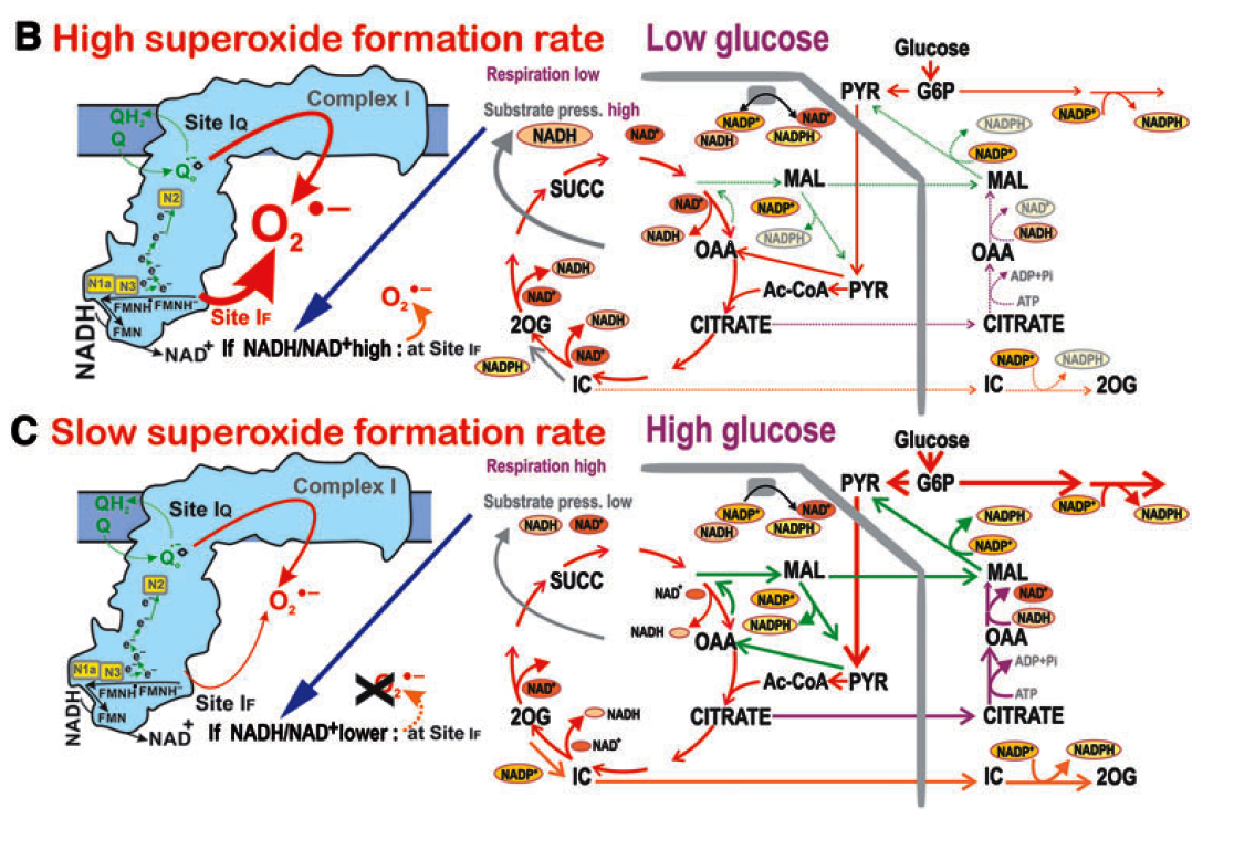
(A) Novel molecular mechanism of insulin secretion emphasizing the role of redox signalization in β cells induced by various substrates (glucose, fatty acids (FA), amino acids (AA), Arginin (Arg)).
(B, C) Overview of major metabolic and redox fluxes in β cell under low, non-stimulating, glucose levels (B, causing no insulin secretion) and under high glucose levels inducing insulin secretion (C).
Plecitá-Hlavatá L, Engstová H, Holendová B, Tauber J, Špaček T, Petrásková L, Křen V, Špačková J, Gotvaldová K,Ježek J, Dlasková A, Smolková K, Ježek P Mitochondrial Superoxide Production Decreases on Glucose-Stimulated Insulin Secretion in Pancreatic beta Cells Due to Decreasing Mitochondrial Matrix NADH/NAD(+) Ratio . Antioxidants & Redox Signaling. 2020; 33(12); 789-815 . IF: 6.323 DOI
Losartan attenuates neuroinflammation and neuropathic pain in paclitaxel-induced peripheral neuropathy (13.10. 2020)
Paclitaxel-induced peripheral neuropathy in patients is often associated with neuropathic pain and neuroinflammation in the central and peripheral nervous system. The analgesic treatments available for this condition have numerous serious side effects and their efficacy is low. There is a great need to understand the neuropathic pain mechanisms involved in order to find new analgesic drugs. Frequently used antihypertensive drug Losartan, an angiotensin II receptor type 1 (AT1R) blocker, was shown to have some anti-inflammatory and neuroprotective effects in different disease models. In our work we have shown that systemic Losartan treatment attenuated significantly mechanical allodynia in a rodent model of Paclitaxel-induced neuropathic pain. Losartan also significantly reduced paclitaxel-induced neuroinflammatory changes and induced expression of pro-resolving markers (Arginase 1 and IL-10) indicating a possible shift in macrophage polarization. Considering the good safety profile of Losartan, it may be considered as a possible novel treatment strategy for patients with paclitaxel induced neuropathic pain.
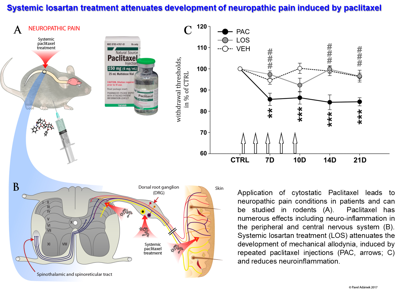
Kalynovska N, Diallo M, Sotáková-Kašparová D, Paleček J Losartan attenuates neuroinflammation and neuropathic pain in paclitaxel-induced peripheral neuropathy. Journal of Cellular and Molecular Medicine 24, 14 (2020), 7949-7958. IF: 4.486 DOI
Signalling bias as the way to side-effect-free medication (22.7. 2020)
More than 30 % of currently marketed medications act via G-protein coupled receptors (GPCRs). This type of membrane receptors is involved in the control of a wide range of physiological processes, from the processing of sensory stimuli, through the regulation of behavior and mood, hormonal and immune responses, to the control of autonomic functions and cell proliferation. Thus, GPCRs represent one of the most important pharmacotherapeutic targets. In contrast to traditional agonists activating multiple signalling pathways, agonists biased towards a given signalling pathway represent a new generation of drugs with increased specificity and fewer adverse effects. Enormous research has been done on agonists biased towards either G-protein or arrestin mediated pathway.
In our laboratory we focus on the study of muscarinic acetylcholine receptors, which belong to the typical representatives of GPCR. Here, as a proof of concept, we demonstrate unprecedented signalling bias solely at the level of G‑protein-mediated signalling. We present agonists of muscarinic acetylcholine receptors, a member of GPCR family, that exclusively inhibit cAMP synthesis through activation of Gi‑protein pathway and thus are functionally selective for M2 and M4 receptor subtypes. Muscarinic receptors M2, M4 represent one of the pharmacological targets in the treatment of pain. Newly discovered agonists may lead to the development of new non-addictive analgesics such as opiates, or immunity-weakening as steroid analgesics. Such muscarinic analgesics would not cause side effects mediated by activation of the Gq protein signaling pathway, such as incontinence, excessive salivation and sweating, and more.
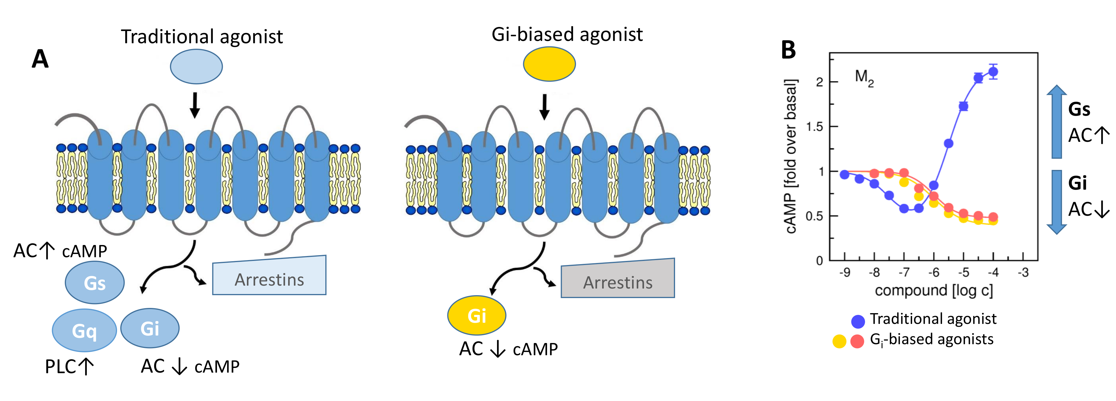
A) General scheme of coupling GPCRs with individual subtypes of G‑proteins or arrestins after activation by a traditional full agonist (left) and an agonist preferring Gi‑protein activation (right). AC, adenylyl cyclase; PLC, phospholipase C. B) Activation of M2 receptor by the traditional agonist (blue) resulting in stimulation of the Gi and Gs‑protein pathway leading to a decrease in cAMP level in the cell at low concentration of agonist and an increase in cAMP level at high concentrations of the agonist. Activation of M2 receptor by two different Gi‑biased agonists (red, orange) resulting in stimulation of the Gi‑protein pathway only and decrease in cAMP level.
Randáková A, Nelic D, Ungerová D, Nwokoye P, Su Q, Doležal V, El-Fakahany EE, Boulos J, Jakubík J. Novel M2-Selective, Gi-Biased Agonists of Muscarinic Acetylcholine Receptors. British Journal of Pharmacology. 2020; 177(9); 2073-2089 . IF: 7.730 DOI
Load next