New findings on the structure of the FOXO4: p53 complex - a key factor in senescence regulation (22.4. 2022)
NEW PUBLICATION
Transcription factor p53 protects cells against tumorigenesis when subjected to various cellular stresses. Under stress conditions, p53 interacts with another transcription factor, FOXO4 (Forkhead box O 4), and together they increase the production of p21 protein, which triggers the process of cell aging (senescence). However, the molecular mechanism of upregulation of p21 transcription is still unclear. In the study published in the Protein Science journal, scientific teams of Dr. Obsilova (IPHYS CAS), prof. Obsil (Faculty of Science, Charles University and IPHYS CAS) and their colleagues from IOCB CAS characterized interactions between p53 and FOXO4 at the molecular level. New knowledge about the structure of the complex may enable the development of specific inhibitors of the interaction between these two proteins, and subsequently in the development of new drugs aimed at the selective elimination of senescent cells.
In this structural study, the researchers performed a detailed characterization of the interactions in the FOXO4: p53 complex using an integrated approach involving analytical ultracentrifugation, nuclear magnetic resonance, and chemical cross-linking coupled to mass spectrometry. Because both FOXO4 and p53 have multiple domains (see Figure), they studied the role of individual domains and disordered segments of both proteins and mapped their interaction interfaces. They found out that the interaction between p53 transactivation domain TAD and the FOXO4 Forkhead domain is crucial for the overall stability of the p53:FOXO4 complex. Furthermore, contacts involving the N-terminal disordered FOXO4 segment, the C-terminal negative regulatory domain of p53, and the DNA-binding domains of both proteins stabilize the complex formation. By measuring DNA binding, they further found that the p53: FOXO4 complex formation blocks p53 binding to DNA without affecting the DNA-binding properties of FOXO4.

Left, sedimentation velocity analytical ultracentrifugation analysis of interaction between FOXO4 and p53. Middle, chemical shift perturbations obtained from 1H-15N HSQC spectra of 15N-labeled FOXO4 in the presence of p53 mapped onto the crystal structure of the FOXO4 DBD:DNA complex. Right, fluorescence anisotropy measurements showing that the complex formation reduces the DNA-binding affinity of p53.
Mandal R, Kohoutova K, Petrvalska O, Horvath M, Srb P, Veverka V, Obsilova V and Obsil T. FOXO4 interacts with p53 TAD and CRD and inhibits its binding to DNA. Protein Sci. roč. 31, č. 5 (2022), č. článku e4287. IF = 6.725. DOI
NEW PUBLICATION
An oral glucose tolerance test (OGTT) is the most commonly used method to diagnose diabetes mellitus from a drop of blood. It measures the ability of an organism to clear circulating glucose after ingestion of glucose bolus after an overnight fast. Although the dynamics of the blood glucose levels during the OGTT are well known, much less information about the metabolic changes in the target organs and the inter-organ communication are available. In our study, we investigated what is the fate of the sugar molecules in each organ and how it affects metabolic pathways in the body. Therefore, we performed the OGTT in mice using glucose with stable isotopic tracers (13C), analyzed 13C6-glucose tissue distribution and time profiles of metabolites and lipids across 12 organs and plasma. We found, that during the OGTT, the glucose use is turned on with specific kinetics at the organ level, but fasting substrates like β-hydroxybutyrate are switched off in all organs simultaneously. Timeline profiling of 13C-labeled fatty acids and triacylglycerols across tissues suggests that brown adipose tissue may contribute to the circulating fatty acid pool at maximal plasma glucose levels. We have created a virtual interactive atlas of metabolites (sugars, amino acids, lipids, etc.), which describes the interactions between organs after ingesting grape sugar. Metabolic fate of ingested glucose carbons was followed in 12 organs and plasma.

Visit the web application to explore virtual mouse metabolome yourself.
Lopes M, Brejchova K, Riecan M, Novakova M, Rossmeisl M, Cajka T, Kuda O. Metabolomics atlas of oral 13C-glucose tolerance test in mice. Cell Rep. 2021 Oct 12;37(2):109833. DOI. PMID: 34644567 IF = 9,423
The effectivity of opioid analgesics is reduced by CCL2 chemokine (8.12. 2021)
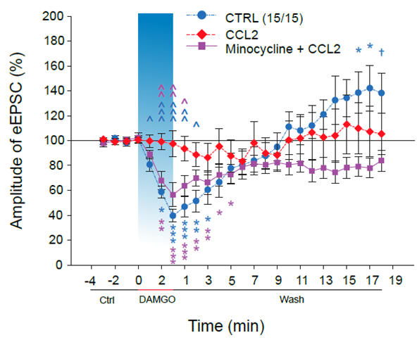
Opioid analgesics are the standard of care in the treatment of serious painful states. Treatment of neuropathic pain states, induced by damage to the nervous system, is especially difficult and opioid analgesics often do not have a beneficial effect. It was shown before that neuropathic states are accompanied with neuroinflammatory changes in the spinal cord and the level of different signaling molecules such as chemokine CCL2 is increased. This work shows that chemokine CCL2 is one of the important factors significantly reducing the effectivity of analgesics acting through opioid receptors. It acts probably directly on neurons and also through activation of microglial cells. Analgesic treatment with opioids has also number of serious unwanted side effects. One of them is a paradoxical increase of sensitivity, hyperalgesia/pain after opioids administration. This work shows that this hyperalgesia may be related to TRPV1 receptors activation. These published results suggest that to improve pain treatment with opioid analgesics, modulation of CCL2 and TRPV1 receptors may be needed, especially in cases of neuropathic pain.

The inhibition of the opioid agonists induced reduction of painful/nociceptive signaling in the spinal cord dorsal horn by the CCL2 chemokine. The control blue line demonstrates the inhibition of the nociceptive synaptic signaling after the DAMGO application and later (in about 13minutes) its potentiation. DAMGO is µ opioid receptors agonist and simulates thus application of opioid analgesics. The analgesic effect of DAMGO application is completely reversed in the presence of CCL2 chemokine (red line). The purple line demonstrates that the effect of CCL2 is dependent on microglia cells activation as it is prevented by microglia inhibitor minocycline.
Chemokine CCL2 preventsopioid‑inducedinhibitionofnociceptivesynaptictransmission in spinalcorddorsalhorn. Mario Heles, Petra Mrozkova, Dominika Sulcova, Pavel Adamek, Diana Spicarova and Jiri Palecek, JournalofNeuroinflammation (2021) 18:279, DOI, IF=8.23
Laboratory of Pain Research, Institute of Physiology, The Czech Academy of Sciences, Videnska 1083, 142 20 Praha 4, Czech Republic
Gliflozins - more than just antidiabetics (8.12. 2021)
Gliflozins are commonly prescribed for the treatment of diabetes (patients may know e.g. empagliflozin under the brand name Jardiance). Gliflozins inhibit the activity of sodium-glucose transporter in kidneys, which leads to higher glucose excretion in urine and normalization of blood glucose levels. Besides alleviation of hyperglycemia, other beneficial effects were observed in patients taking gliflozins, including decreased body weight, reduced blood pressure or improved kidney function. How this is possible? Here comes the right time to take a step backwards and to look once again and in greater detail on gliflozins effects in rodent models. And that is exactly what dr. Vaneckova and her colleagues from IKEM and departments of IPHYS are doing. They characterized the effects of empagliflozin in various non-diabetic rodent models prone to hypertension (Ren-2 transgenic rats; TGR) and lipid imbalance (hereditary hypertensive rats). Although the details of treatment effects vary among the individual models, empagliflozin generally attenuated inflammation, and normalized plasma and tissue lipid levels. In addition, it inhibited the activity of sympathetic nervous system, which resulted in a decrease of blood pressure in TGR. Hopefully, this and similar findings will allow to extend the use of gliflozins in the future to more diagnoses than just diabetes.


Hojná S, Rauchová H, Malínská H, Marková I, Hüttl M, Papoušek F, Behuliak M, Miklánková D, Vaňourková Z, Neckář J, Kadlecová M, Kujal P, Zicha J, Vaněčková I. Antihypertensive and metabolic effects of empagliflozin in Ren-2 transgenic rats, an experimental non-diabetic model of hypertension. Biomed Pharmacother. 2021 Dec;144:112246. Epub 2021 Oct 1. IF-5,98, DOI
Targeted modulation of NMDA receptors is a key for the effective treatment of neurological and neurodevelopmental diseases (14.10. 2021)
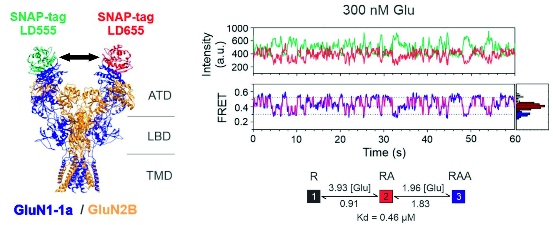
N-methyl-D-aspartate (NMDA) receptors are glutamate-gated ion channels critically involved in excitatory synaptic transmission that play a key role in learning and memory. Impaired NMDA receptor function leads to major neurological, neurodevelopmental and psychiatric disorders such as schizophrenia, autistic spectrum disorder, epilepsy or Alzheimer's disease. For an effective design of novel drugs capable of specifically modulating NMDA receptors, it is essential not only to understand the NMDA receptor atomic structure, but also to uncover the specific sequence of conformational changes that are involved in receptor activation and allosteric modulation.
We used single-molecule FRET to identify and quantify the sequence of conformational changes in the amino-terminal domain of the NMDA receptor during its activation. Next, we uncovered distinct roles of receptor subunits in receptor activation, and last but not least, we have identified the molecular mechanism of receptor modulation by pH during pathophysiological conditions such as stroke.

Vyklický, Vojtěch - Stanley, Ch. - Habrian, Ch. - Isacoff, E. Y. Conformational rearrangement of the NMDA receptor amino-terminal domain during activation and allosteric modulation. Nature Communications. Roč. 12, č. 1 (2021), č. článku 2694. ISSN 2041-1723. E-ISSN 2041-1723, IF: 14.919, rok: 2020, DOI
Structural basis of heat-induced opening of TRP channels (12.10. 2021)
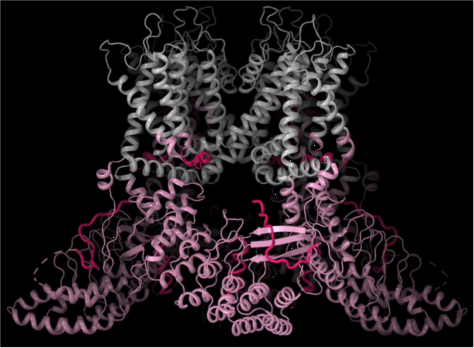
TRPV3 is an ion channel involved in the detection of temperature changes, pain, itching, skin barrier maintenance, wound healing and hair growth. Disturbances in its function are the cause of many serious human skin diseases, including the genodermatosis known as Olmsted syndrome, atopic dermatitis, rosacea and psoriasis. An international team of scientists led by Prof. Alexander Sobolevsky (Columbia University, New York, NY, USA), in collaboration with scientists from the Institute of Physiology of the Czech Academy of Sciences in Prague, has identified how the TRPV3 channel is altered by heat and determined the molecular basis of its activation. The study shows that opening of TRPV3 by temperatures exceeding the physiological threshold (higher than 40 °C) involves changes in the secondary structure of specific regions of the channel protein complex, but also active participation of membrane lipids. The result is an important contribution to the understanding of the general molecular mechanisms of thermal activation of TRPV channels and a prerequisite for the search for possible approaches to their pharmacological regulation. The importance of the research on these remarkable protein complexes is evidenced by the recent award of the 2021 Nobel Prize in Physiology or Medicine to two American scientists, David Julius and Ardem Patapoutian.


Left: transition between open and closed states of TRPV3 subjected to temperature cycles. Structures were obtained by Cryo-EM. Right, structural transitions between closed, sensitized and open states. Dynamic regions are highlighted in pink, the elements undergoing the strongest structural changes are highlighted in dark pink.
Nadezhdin, K. D. - Neuberger, A. - Trofimov, Yu. A. - Krylov, N. A. - Sinica, Viktor - Kupko, N. - Vlachová, Viktorie - Zakharian, E. - Efremov, R. G. - Sobolevsky, A. I. Structural mechanism of heat-induced opening of a temperature-sensitive TRP channel. Nature Structural & Molecular Biology. Roč. 28, č. 7 (2021), s. 564-572. ISSN 1545-9993. E-ISSN 1545-9985, IF: 15.369, rok: 2020, DOI
New findings may help to improve diagnostic methods for breast cancer (23.8. 2021)
2-hydroxyglutarate (2HG) is a metabolite resembling normal cell metabolite 2-oxoglutarate (2OG), however, its accumulation in cells might lead to amplification of processes in cancer development. R-2HG is a product or bi-product of several metabolic enzymes, including mitochondrial ones. We investigated whether production of mitochondrial 2HG is elevated in breast cancer cell lines and identified active competition for initial substrate, 2OG, between enzymes isocitrate dehydrogenase IDH2 and alcohol dehydrogenase ADHFE1. We have also investigated possible substrate and cofactor NADPH channeling between the two IDH2 molecules within mitochondria. We characterized several situations when either IDH2 and ADHFE1 produce a non-negligible amount of 2HG, which is then actively exported from cells. This can serve as a basis for clinical application of our findings. We have therefore quantified 2HG levels in the urine of breast carcinoma patients after resection of their tumors and showed a positive correlations between cancer stages and 2HG levels. Note that cancer stages I to IV differ by the existence, localization and severity of metastases. A future extension of these findings might help to improve diagnostic approaches of breast carcinoma.
Špačková; Jitka - Gotvaldová; Klára - Dvořák; Aleš - Urbančoková; Alexandra - Pospíšilová; K. - Větvička; D. - Leguina-Ruzzi; Alberto A. - Tesařová; P. - Vítek; L. - Ježek; Petr - Smolková; Katarína . Biochemical Background in Mitochondria Affects 2HG Production by IDH2 and ADHFE1 in Breast Carcinoma . Cancers (Basel). 2021; 13(7)); 1709 . IF = 6.639 [ASEP] [ DOI ]

Palmitoylation controls NMDA receptor susceptibility to neurosteroids (3.5. 2021)
N-methyl-D-aspartate (NMDA) receptors are ionotropic glutamate receptors that are crucial for synaptic transmission, learning, and memory acquisition. Their overactivation leads to pathology associated, for example, with stroke or Alzheimer's disease. Overactivation of NMDA receptors can be inhibited by a number of substances, including neurosteroids.
Using electrophysiological and molecular-biological techniques, we have elucidated the molecular mechanism by which NMDA receptor susceptibility to inhibitory neurosteroids is increased. This change is due to depalmitoylation of three cysteines (C849, C854, C871) in the intracellular part of the GluN2B receptor subunit, which occurs after a transient increase in intracellular concentration of Ca2+. Beyond the pharmacological consequences, depalmitoylation of the receptor results in a change in kinetic parameters in favor of the closed state. Increased sensitivity of NMDA receptors to inhibitory neurosteroids is thus another of the neuroprotective mechanisms that prevents excitotoxic damage to nerve tissue.
Hubálková, Pavla - Ladislav, Marek - Vyklický, Vojtěch - Smejkalová, Tereza - Hrčka Krausová, Barbora - Kysilov, Bohdan - Krůšek, Jan - Naimová, Žaneta - Kořínek, Miloslav - Chodounská, Hana -Kudová, Eva - Černý, Jiří - Vyklický ml., Ladislav Palmitoylation Controls NMDA Receptor Function and Steroid Sensitivity. Journal of Neuroscience 2021. 41 (10) 2119-2134, F: 5.674 DOI

Clove oil alleviates cold-induced toothache by blocking the TRPC5 ion channel (30.4. 2021)
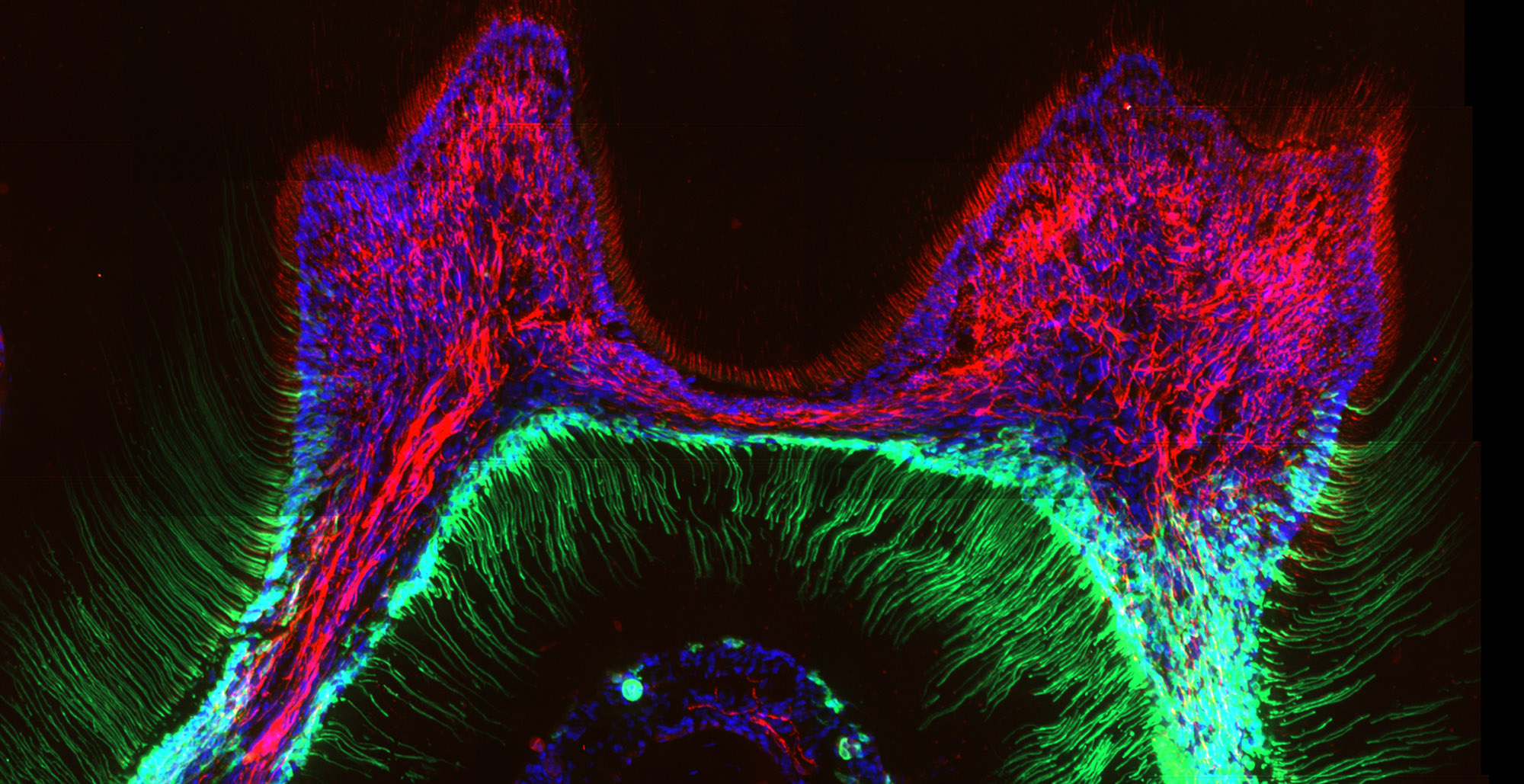
Touching a cold drink can be a suffering for us when we have tooth decay. An international team of scientists led by Prof. Katharina Zimmermann (Friedrich - Alexander University Erlangen - Nuremberg in Germany), together with scientists from the Institute of Physiology of the Czech Academy of Sciences in Prague, found out how teeth detect cold and determined the molecular basis of cold-induced dental pain. In both mice and humans, dental cells called odontoblasts contain special cold sensors, the TRPC5 ion channels, which transmit information about a painful stimulus to the nervous system. The study also offers an explanation for why clove oil, which has been used for centuries in dentistry, can alleviate toothache. Clove oil contains a chemical eugenol that blocks the TRPC5 protein and prevents it from activating nerves. Video

Odontoblasts containing the ion channel TRPC5 (green) tightly pack the area between the pulp and the dentin in a mouse’s molar. The cells’ long-haired extensions fill the thin canals in dentin that extend towards the enamel. Sensory nerves are indicated in red (bIII-tubulin). Cell nuclei are stained with Hoechst 33258. (Credit: L. Bernal et al./Science Advances 2021)
Bernal, L. - Sotelo-Hitschfeld, P. - König, Ch. - Sinica, Viktor - Wyatt, A. - Winter, Z. - Hein, A. - Touška, Filip - Reinhardt, S. - Tragl, A. - Kusuda, R. - Wartenberg, P. - Sclaroff, A. - Pfeifer, J. D. -Ectors, F. - Dahl, A. - Freichel, M. - Vlachová, Viktorie - Brauchi, S. - Roza, C. - Boehm, U. - Clapham, D. E. - Lennerz, J. K. - Zimmermann, K. Odontoblast TRPC5 channels signal cold pain in teeth. Science Advances. Roč. 7, č. 13 (2021), č. článku eabf5567. ISSN 2375-2548. IF: 13.117, rok: 2019. DOI
Nanocellulose as a promising cell carrier for applications in regenerative medicine (8.4. 2021)
Cellulose in the form of a fabric has been used for thousands of years as a traditional wound dressing material. Nowadays, it can be used not only for passive wound covering, but also for active wound healing, e.g. with controlled delivery of various drugs or cells for regenerating the damaged tissue. A suitable form of cellulose for this purpose is nanocellulose, i.e. cellulose in the form of nanofibrils, simulating the architecture of the native extracellular matrix. This form of cellulose is produced by some species of bacteria, or can be isolated from higher plants, including wood. In our experiments, we have focused on the development of “intelligent” wound dressings, capable of delivering skin and stem cells into skin wounds. These dressings are based on electrically-charged cellulose nanofibrils, attached to a microfibrous cellulose fabric. Anionic nanocellulose provided a suitable substrate for the adhesion and growth of human dermal fibroblasts and human adipose tissue-derived stem cells, while cationic nanocellulose provided better support for cell-cell adhesion and for the formation of cell aggregates, which was apparent mainly in fibroblasts (Fig. 1). This difference was due to the preferential adsorption of albumin from the serum supplement of the culture medium, which is non-adhesive for cells, on cationic nanocellulose. However, both types of nanocellulose are useful in regenerative medicine. Anionic nanocellulose is suitable for creating continuous cell sheets, which can be delivered into the wound either spontaneously or after release from the substrate using cellulase enzymes, while cationic cellulose is suitable for creating cell spheroids, i.e. important structures for developing organoids and for tissue engineering.

Fig. 1 Morphology of normal human dermal fibroblasts (NHDFs; A) and human adipose tissue-derived stem cells (ADSCs; B), guided by the topography of cellulose meshes coated with anionic or cationic cellulose nanofibrils (a600, c600, respectively) after seven days of cultivation. 3D projection of microscopy images (front view and side view) of the cells on the material. F-actin of the cell cytoskeleton is stained in red, vinculin in the cells is stained in green. Confocal microscope with objective magnification 40x.
Pajorova J, Skogberg A, Hadraba D, Broz A, Travnickova M, Zikmundova M, Honkanen M, Hannula M, Lahtinen P, Tomkova M, Bacakova L, Kallio P. A cellulose mesh with charged nanocellulose coatings as a promising carrier of skin and stem cells for regenerative applications. Biomacromolecules, 2020, 21: 4857-4870, https://dx.doi.org/10.1021/acs.biomac.0c01097; IF = 6.092; DOI
Load next


