Heat or cold? New insights into the functioning of temperature-sensitive ion channels obtained in collaboration with Lund University (24.1. 2023)
NEW PUBLICATION
Temperature-sensitive TRP ion channels are cellular molecular sensors involved in the transduction of sensory signals, pain perception (nociception) and the maintenance of ion homeostasis. Disturbances in their function are associated with many serious human diseases such as chronic pain, inflammation, cancer and various cardiovascular, neurological, respiratory, renal and metabolic disorders. The enormous hope of developing new drugs targeting these channels is greatly limited due to their intrinsic ability to be activated by stimuli of different modalities (polymodality), the mechanism of which has not yet been fully understood. An international team of scientists led by Prof. Peter M. Zygmund (Lund University, Malmö, Sweden), in collaboration with scientists from the Institute of Physiology of the Czech Academy of Sciences in Prague, has revealed two important mechanisms contributing to the polymodal regulation of TRP channels: The first study revealed the binding site and molecular nature of the binding of D9‑tetrahydrocannabiorcol (a natural plant cannabinoid with no psychotropic effects), which strongly facilitates the activation of TRPV2, thereby affecting the transmission of painful stimuli. The second study identified two separate specific regions that confer heat (> 35 °C) and cold (< 15 °C) sensitivity to a different receptor, TRPA1, and demonstrated that the temperature sensitivity of this receptor is critically dependent on oxidative and reducing environments. The results make an important contribution to the understanding of the general molecular mechanisms of chemical and thermal activation of TRP family channels and will find application in the search for possible approaches to their regulation by drugs.

Left panel: effect of cold (15°C) on activation of the human TRPA1 wild-type ion channel and a mutant in which the major amino acid residue C856 responsible for regulation by oxidizing and reducing agents is substituted for alanine. The channels were activated by a series of voltage pulses (from ‑160 mV to +200 mV). Right panel: visualization of the TRPA1 channel structure obtained by cryoelectron microscopy (PDB: 6v9w) as viewed from the side and top. Dynamic regions of the channel responsible for cold activation are highlighted in light blue. The position of cysteine C856 is shown in the detail of the structure of one subunit below.
Moparthi, L. - Sinica, Viktor - Moparthi, V. K. - Kreir, M. - Vignane, T. - Filipovic, M. R. - Vlachová, Viktorie - Zygmunt, P. M.The human TRPA1 intrinsic cold and heat sensitivity involves separate channel structures beyond the N-ARD domain. Nature Communications. Roč. 13, č. 1 (2022), IF: 17.694 DOI
Zhang, L. - Simonsen, Ch. - Zímová, Lucie - Wang, K. - Moparthi, L. - Gaudet, R. - Ekoff, M. - Nilsson, G. - Hellmich, U. A. - Vlachová, Viktorie - Gourdon, P. - Zygmunt, P. M. Cannabinoid non-cannabidiol site modulation of TRPV2 structure and function. Nature Communications. Roč. 13, č. 1 (2022), IF: 17.694, rok: 2021 DOI
Unravelling novel regulatory mechanisms in the human Na+/H+ antiporter NHA2 (14.11. 2022)
Sodium/proton antiporters are membrane proteins found in all living cells from bacteria to humans and play an important role in regulating intracellular pH and cation concentrations. Among these types of transporters belong also the NHA2 antiporter, whose activity affects a number of physiological functions, e.g. insulin secretion, sodium reabsorption in the kidneys or sperm motility. The NHA2 antiporter transports Na+ or Li+ cations across the membrane in exchange for H+ and its activity is specifically inhibited by phloretin. The properties and functions of all proteins result from their primary structure, i.e. from the sequence of amino acids from which the protein consists. In the family of Na+/H+ antiporters, NHA2 has a unique structure. It consists of 537 amino acids with 14 transmembrane domains and an unique hydrophilic N-terminus long 82 amino acid residues, whose structure and function have not yet been studied. Our new results published in the Protein Science journal revealed several new structural elements important for the NHA2’s function, including the unravelling a new regulatory role of the hydrophilic N-terminus.
We studied the human NHA2 protein and its mutated variants using its expression in a model eukaryotic organism, the yeast Saccharomyces cerevisiae, as well as using bioinformatic simulations (in collaboration with the laboratory of Prof. Nir Ben-Tal from Tel-Aviv University). We newly identified several amino acid residues important for antiporter selectivity (recognition and transport of Na+ and Li+ cations) and for transport of protons. Furthermore, we determined the place in the protein structure where the phloretin inhibitor binds. We also revealed that the unique hydrophilic N-terminal part of the protein has an important (autoinhibitory) role in regulating the transport activity of NHA2, because truncations of the first 50 - 70 residues of the N-terminus doubled the transport activity of the antiporter. Our results also show that the new expression system for NHA2 in yeast cells can be useful for a rapid screening of SNP´s effects on NHA2 activity, and/or to test new compounds influencing the function of NHA2, similarly as phloretin.

Velazquez D., Prusa V., Masrati G., Yariv E., Sychrova H., Ben-Tal N. and Zimmermannova O. (2022): Allosteric links between the hydrophilic N-terminus and transmembrane core of human Na+/H+ antiporter NHA2. Protein Sci: e4460. IF = 6.993 DOI
Thiazolidinediones (TZDs) belong to the category of antidiabetic drugs that increase insulin sensitivity. However, the first generation of TZDs show several side effects, including increased formation of large fat cells (adipocytes) in the bone marrow associated with a higher risk of fractures and bone loss. These side effects are thought to be caused by the strong binding of TZDs to the nuclear receptor PPARγ. In a new study published in the journal Molecular Metabolism, we investigated how administration of MSDC-0602K, a new TZD analog with lower affinity for PPARγ receptors, affects bone metabolism.
We found that administration of MSDC-0602K to obese mice for 8 weeks resulted in significantly better bone microstructure and bone strength, along with an increased proportion of smaller adipocytes in the bone marrow, compared to the original TZDs. We also investigated the effects of MSDC-0602K at the molecular level using primary cell cultures. Bone marrow mesenchymal stem cells from mice treated with MSDC-0602K differentiated to an increased extent into the form of osteoblasts (bone cells), did not undergo as much aging and showed increased cellular metabolism of glutamine, which is important for bone formation (osteogenesis).
Taken together our findings suggests that the new TZD analog could increase insulin sensitivity with less adverse effects on bone quality and mesenchymal stem cell metabolism compared to the original TZDs. Thus, MSDC-0602K could replace older antidiabetic drugs in the treatment of metabolic and bone diseases.

Benova, A., M. Ferencakova, K. Bardova, J. Funda, J. Prochazka, F. Spoutil, T. Cajka, M. Dzubanova, T. Balcaen, G. Kerckhofs, W. Willekens, G. H. van Lenthe, G. Alquicer, A. Pecinova, T. Mracek, O. Horakova, M. Rossmeisl, J. Kopecky and M. Tencerova (2022). "Novel thiazolidinedione analog reduces a negative impact on bone and mesenchymal stem cell properties in obese mice compared to classical thiazolidinediones." Mol Metab 65: 101598. IF = 8.568 DOI
The structure of steroids fundamentally affects their effect at the N-methyl-D-aspartate receptors (20.10. 2022)
N-methyl-D-aspartate receptors (NMDARs) are proteins involved in the regulation of many processes in the mammalian brain. They play a key role in signal transmission between nerve cells, and even a small disturbance in the NMDAR function may have sever pathophysiological effects. A reduced function of NMDARs is associated with neuropsychiatric disorders such as mental retardation, reduced intellect, schizophrenia, autism spectrum disorders, epilepsy, or motor disorders. The insufficient NMDAR function can be compensated for by numerous compounds, including neurosteroids. However, the mechanism by which neurosteroids potentiate NMDARs is not well understood.
In a study published in the British Journal of Pharmacology, we investigated the effect of newly synthesized synthetic analogues of endogenous neurosteroid pregnanolone sulfate on NMDARs. We demonstrated that analogues with short aliphatic chains, such as pregnanolone carboxylate (PA-Car), inhibit NMDAR responses, whereas analogues with longer aliphatic chains, such as epipregnanolone butyrate (EPA-But), potentiate NMDAR responses. Combining electrophysiology, molecular biology, and computational modelling, we identified the binding site for EPA-But at the transmembrane domain of NMDAR and suggested the mechanism by which EPA-But enhance the NMDAR function.

(A) Left, structure of pregnanolone carboxylate (PA-Car) and epipregnanolone butyrate (EPA-But). Right, graph shows the average effect of PA-Car and EPA-But at NMDAR. (B) Location of the EPA-But binding site at the transmembrane domain of NMDAR. Amino-acid residues that are involved in the interaction with EPA-But molecule (green) are highlighted in red.
Kysilov B, Hrcka Krausova B, Vyklicky V, Smejkalova T, Korinek M, Horak M, Chodounska H, Kudova E, Cerny J, Vyklicky L: Pregnane-based steroids are novel positive NMDA receptor modulators that may compensate for the effect of loss-of-function disease-associated GRIN mutations. British Journal of Pharmacology, (2022) 179:15, 3970–3990, IF = 9.473, DOI
Ion channel TRPC5 as a target for treating neuropathic pain (19.10. 2022)
Despite great advances in medicine, treatment options for pain states associated with diabetes or chemotherapy-induced neuropathy are still limited. These types of neuropathies are accompanied by cold-induced pain that is very difficult to manage in clinical practice. In fact, the only drug that has successfully undergone clinical trials and demonstrated efficacy for this type of pain so far is duloxetine. This drug is superior to other antidepressants with a similar mechanism of action at managing painful neuropathies, but it is not known why. Interestingly, it has recently been shown that the cold-sensitive TRPC5 ion channel is expressed in human sensory neurons and that inhibition of its activity relieves persistent pain, including neuropathic cold pain.
In our study we asked whether the TRPC5 channel is modulated by duloxetine and may contribute to its analgesic effect. Our electrophysiological measurements showed that TRPC5 channel activity is strongly suppressed by duloxetine. We performed molecular docking and molecular dynamic simulations that identified a potential biding site for duloxetine. Subsequent point mutagenesis validated that the duloxetine molecule resides in a well-known biding pocket on the intracellular side of the TRPC5 transmembrane domain. Slight manipulation of the shape and electrostatic of the binding pocket environment (replacing the amino acid glutamate 418 with alanine) caused a complete loss of the duloxetine effect on voltage-evoked TRPC5 activity. Our results suggest that TRPC5 is a previously unrecognised target for a commonly used, highly effective drug against severe forms of pain. Furthermore, the finding that this TRPC5 inhibitor is widely used and well tolerated provides a scaffold for new pain treatment strategies.

Zimova L, Ptakova A, Mitro M, Krusek J,Vlachova V: Activity dependent inhibition of TRPC1/4/5 channels by duloxetine involves voltage sensor-like domain, Biomedicine & Pharmacotherapy. Roč. 152, August 2022, 113262, IF = 7,419, DOI
New lipid classes in human breastmilk (29.7. 2022)
Breastfeeding is the best way of nutrition for a newborn baby. Mother's milk provides the ideal cocktail of nutrients and bioactive components for rapid growth and development in this early period of human life. Milk composition can be affected by several factors such as maternal dietary habits, mode of delivery, and pregnancy length. An essential question for neonatologists is just the optimal composition of breastmilk for premature babies.
In our study, we investigated the impact of preterm birth and a cesarean section on the quality of the first form of milk – colostrum. Analysis of the samples obtained from mothers at the Institute for the Care of Mother and Child in Prague showed that both the mode and term of delivery slightly negatively affected the composition of colostrum. In these cases, the mammary gland of mothers did not have enough time to initiate fully-fledged milk production. Still, this negative effect disappears in time, and mature milk composition resembles the milk of mothers who delivered spontaneously in term.
Moreover, we discovered a peculiar class of lipids called triacylglycerol estolides in all milk samples, which serves as a reservoir of the antiinflammatory lipids. We found out that the milk enzyme carboxyl ester lipase can digest these complex molecules and release substances with beneficial effects on the health of newborns.

Brejchová K, Palůchová V, Březinová M, Čajka T, Balas L, Durand T, Křížová M, Straňák Z, Kuda O: Triacylglycerols containing branched palmitic acid ester of hydroxystearic acid (PAHSA) are present in the breast milk and hydrolyzed by carboxyl ester lipase, Food Chemistry. 2022; 388(Sep 15)); 132983, IF = 9.231 DOI
Maternal rhythmic behavior supports the development of the fetal biological clock (27.5. 2022)
Before the fetal internal biological clock in the suprachiasmatic nuclei (SCN) of the hypothalamus begins to tick, the rhythmic behavior of the mother affects the function and development of this structure. This was found in a new study published by the team of Alena Sumová from the Institute of Physiology of the Academy of Sciences of the Czech Republic on May 24 in the journal PLOS Biology. This discovery contributes significantly to the understanding of the development of the internal clock and may find application in the treatment of premature babies.
The SCN are the central clock in our body, driven by the rhythmic switching on and off of the clock genes. The rhythmic activity of clock genes in SCN cells controls the activity of many other genes locally and elsewhere in the body, which ultimately affects a wide range of circadian rhythms in behavior, including food intake and sleep. However, this autonomous rhythmic gene activity of the SCN begins relatively late in fetal development, which raises the question of whether maternal signals may affect the gene activity in the SCN before it develops.
To investigate this question, the authors compared the daily gene activity profiles in SCN from fetuses developing in pregnant rats kept in the constant darkness under two different conditions. The control group of mothers had intact SCN and free access to food, while the other group of mothers had SCN surgically removed and had limited access to food for eight hours a day so that their locomotor activity showed a circadian rhythm even in the absence of a central clock. Using biostatistical analyzes they found that in the SCN of the fetuses of both groups, there was a small set of genes whose timing differed between the two groups, and a much larger set of genes whose activity oscillated in synchrony. Many of these genes could be assigned to two major neuronal processes, reflecting in the first case the ongoing development of the SCN and in the second case the earliest manifestation of their function. The data suggest that in the development of fetal SCN, maternal stimuli can replace the missing intercellular synaptic communication and control the rhythms of cell populations before the SCN clocks fully mature. The unexpected extent and specificity of SCN cell responses to maternal signals underlines the importance of a functioning maternal biological clock during pregnancy and points to the potential impact of the absence of such signals in preterm infants.

Greiner P., Houdek P., Sládek M., Sumová A.: Early rhythmicity in the fetal suprachiasmatic nuclei in response to maternal signals detected by omics approach. PLOS Biology; DOI, IF 8.029 Published: May 24, 2022
New findings on the structure of the FOXO4: p53 complex - a key factor in senescence regulation (22.4. 2022)
Transcription factor p53 protects cells against tumorigenesis when subjected to various cellular stresses. Under stress conditions, p53 interacts with another transcription factor, FOXO4 (Forkhead box O 4), and together they increase the production of p21 protein, which triggers the process of cell aging (senescence). However, the molecular mechanism of upregulation of p21 transcription is still unclear. In the study published in the Protein Science journal, scientific teams of Dr. Obsilova (IPHYS CAS), prof. Obsil (Faculty of Science, Charles University and IPHYS CAS) and their colleagues from IOCB CAS characterized interactions between p53 and FOXO4 at the molecular level. New knowledge about the structure of the complex may enable the development of specific inhibitors of the interaction between these two proteins, and subsequently in the development of new drugs aimed at the selective elimination of senescent cells.
In this structural study, the researchers performed a detailed characterization of the interactions in the FOXO4: p53 complex using an integrated approach involving analytical ultracentrifugation, nuclear magnetic resonance, and chemical cross-linking coupled to mass spectrometry. Because both FOXO4 and p53 have multiple domains (see Figure), they studied the role of individual domains and disordered segments of both proteins and mapped their interaction interfaces. They found out that the interaction between p53 transactivation domain TAD and the FOXO4 Forkhead domain is crucial for the overall stability of the p53:FOXO4 complex. Furthermore, contacts involving the N-terminal disordered FOXO4 segment, the C-terminal negative regulatory domain of p53, and the DNA-binding domains of both proteins stabilize the complex formation. By measuring DNA binding, they further found that the p53: FOXO4 complex formation blocks p53 binding to DNA without affecting the DNA-binding properties of FOXO4.

Left, sedimentation velocity analytical ultracentrifugation analysis of interaction between FOXO4 and p53. Middle, chemical shift perturbations obtained from 1H-15N HSQC spectra of 15N-labeled FOXO4 in the presence of p53 mapped onto the crystal structure of the FOXO4 DBD:DNA complex. Right, fluorescence anisotropy measurements showing that the complex formation reduces the DNA-binding affinity of p53.
Mandal R, Kohoutova K, Petrvalska O, Horvath M, Srb P, Veverka V, Obsilova V and Obsil T. FOXO4 interacts with p53 TAD and CRD and inhibits its binding to DNA. Protein Sci. roč. 31, č. 5 (2022), č. článku e4287. IF = 6.725. DOI
An oral glucose tolerance test (OGTT) is the most commonly used method to diagnose diabetes mellitus from a drop of blood. It measures the ability of an organism to clear circulating glucose after ingestion of glucose bolus after an overnight fast. Although the dynamics of the blood glucose levels during the OGTT are well known, much less information about the metabolic changes in the target organs and the inter-organ communication are available. In our study, we investigated what is the fate of the sugar molecules in each organ and how it affects metabolic pathways in the body. Therefore, we performed the OGTT in mice using glucose with stable isotopic tracers (13C), analyzed 13C6-glucose tissue distribution and time profiles of metabolites and lipids across 12 organs and plasma. We found, that during the OGTT, the glucose use is turned on with specific kinetics at the organ level, but fasting substrates like β-hydroxybutyrate are switched off in all organs simultaneously. Timeline profiling of 13C-labeled fatty acids and triacylglycerols across tissues suggests that brown adipose tissue may contribute to the circulating fatty acid pool at maximal plasma glucose levels. We have created a virtual interactive atlas of metabolites (sugars, amino acids, lipids, etc.), which describes the interactions between organs after ingesting grape sugar. Metabolic fate of ingested glucose carbons was followed in 12 organs and plasma.

Visit the web application to explore virtual mouse metabolome yourself.
Lopes M, Brejchova K, Riecan M, Novakova M, Rossmeisl M, Cajka T, Kuda O. Metabolomics atlas of oral 13C-glucose tolerance test in mice. Cell Rep. 2021 Oct 12;37(2):109833. DOI. PMID: 34644567 IF = 9,423
The effectivity of opioid analgesics is reduced by CCL2 chemokine (8.12. 2021)
Opioid analgesics are the standard of care in the treatment of serious painful states. Treatment of neuropathic pain states, induced by damage to the nervous system, is especially difficult and opioid analgesics often do not have a beneficial effect. It was shown before that neuropathic states are accompanied with neuroinflammatory changes in the spinal cord and the level of different signaling molecules such as chemokine CCL2 is increased. This work shows that chemokine CCL2 is one of the important factors significantly reducing the effectivity of analgesics acting through opioid receptors. It acts probably directly on neurons and also through activation of microglial cells. Analgesic treatment with opioids has also number of serious unwanted side effects. One of them is a paradoxical increase of sensitivity, hyperalgesia/pain after opioids administration. This work shows that this hyperalgesia may be related to TRPV1 receptors activation. These published results suggest that to improve pain treatment with opioid analgesics, modulation of CCL2 and TRPV1 receptors may be needed, especially in cases of neuropathic pain.
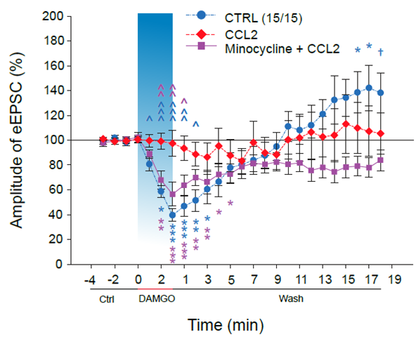
The inhibition of the opioid agonists induced reduction of painful/nociceptive signaling in the spinal cord dorsal horn by the CCL2 chemokine. The control blue line demonstrates the inhibition of the nociceptive synaptic signaling after the DAMGO application and later (in about 13minutes) its potentiation. DAMGO is µ opioid receptors agonist and simulates thus application of opioid analgesics. The analgesic effect of DAMGO application is completely reversed in the presence of CCL2 chemokine (red line). The purple line demonstrates that the effect of CCL2 is dependent on microglia cells activation as it is prevented by microglia inhibitor minocycline.
Chemokine CCL2 preventsopioid‑inducedinhibitionofnociceptivesynaptictransmission in spinalcorddorsalhorn. Mario Heles, Petra Mrozkova, Dominika Sulcova, Pavel Adamek, Diana Spicarova and Jiri Palecek, JournalofNeuroinflammation (2021) 18:279, DOI, IF=8.23
Laboratory of Pain Research, Institute of Physiology, The Czech Academy of Sciences, Videnska 1083, 142 20 Praha 4, Czech Republic
Load next
