

© 2004 - 08 Optical Lab, IBOT, AS CR
 cesky
cesky Academy of Sciences of the Czech Republic
 Institute of Botany, AS CR
Current Info:
• January 2008 •
We apologize for the temporarily incomplete English version of our internet presentation.
 The Main Fields of Interest of the Optical Lab
Microscopic Techniques: Light and electron microscopy form the basis of nearly all the activities in our optical laboratory.
The installed optical (light) microscopes Olympus, with wide field of view, ample depth of field and outstanding performance quality, enables observation and capturing of digital images with magnification range from 3,5× to 1000× (from that: 3,5× till 144× is the continuous range of our stereomicroscope and the discrete values of magnification between 40× and 1000× belong to our “large” microscopes). The “large” research and system microscopes offer beside the brightfield and the darkfield observation/capturing method, the phase contrast, Nomarski differential interference contrast (DIC) or the relief contrast in case of the inverted microscope and separate or simultaneous usage of fluorescence method as well.
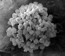
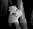
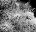
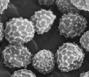
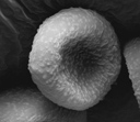
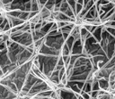
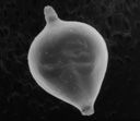
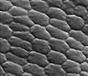
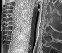
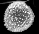
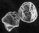
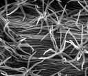
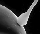
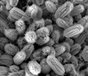
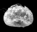
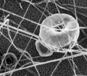
Electron microscopy is in our lab represented by the scanning electron microscope Quanta, made by the FEI Company, and its accessory equipment, especially the sample cooling system. The microscope offers resolution up to 3.5 nm, magnification between 6 and 1 million times and three vacuum modes: High Vacuum, Low Vacuum and the special environmental mode ESEM (the further description will be presented hereinafter).
Digital Capturing: The lab is focusing principally on the digital microphotography, which is tied together with the above-mentioned microscopic techniques. But this area is covered also with several supplementary activities, as is the scanning of negatives and slides, the large format scanning (up to A3 in size, including transparent matters) and the digital photography, mostly the macro photography.
All of the microscopes in our lab after connecting them to our special digital equipment are applicable to observation, editing and capturing of a live image directly on the large LCD of the dedicated work station.
The maximum picture size is 11 megapixels in case of the electron microscope, and 12.5 megapixels in case of the light microscopes. Our compact digital cameras offer at most 5 mega pixel pictures, the optical resolution of the flatbed scanner is 1400×2800 dpi and its maximum resolution with software interpolation is 9600×9600 dpi.
Digital Image Processing and Analysis: The Optical Lab software equipment enables further processing and analysis of the digitalized image, including the structure enhancement (e.g. visualisation of chromosomes), image transformation, calibration together with application of various measurements and/or special calculations, etc.
 Scanning Electron Microscopy
Scanning Electron Microscope
ESEM (Environmental Scanning Electron Microscope)
Apology: The English version of these paragraphs is not available for the time being. Please try it later or see the Czech version.

Reviewed by: Jiri Machac, 8. 1. 2008.
First published: 12. 2. 2007. Last updated: 8. 1. 2008. Developed and maintained by: Jiri Machac.
Copyright © 2004 - 08 Institute of Botany, Academy of Sciences of the Czech Republic