|
 |
|

|
|
|
Mouse
embryonal fibroblast 3T3. Interphase cell.
|
|
Mouse embryonal fibroblast 3T3. Metaphase,
anaphase and telophase.
|
|
Tubulin stained with polyclonal antibody (green),
gamma-tubulin stained with antibody TU-30 (red), DNA (blue). |
|
Tubulin stained with polyclonal antibody (green),
gamma-tubulin stained with antibody TU-30 (red), DNA (blue).
|
|
|
|
|
|
|

|
|

|
|
|
Mouse embryonal fibroblast 3T3.
|
|
Mouse embryonal fibroblast 3T3 treated with anti-mitotic drug
vinblastine.
|
|
|
Cells were microinjected with antibody TU-07 recognizing epitope
within C-terminal structural domain of alpha-tubulin (green). Preparation was
thereafter fixed and stained with polyclonal antibody against tubulin
(red) and DNA-binding dye (blue).
|
|
Tubulin paracrystals stained with antibody TU-01
against alpha-tubulin (red), vimentin coils stained with antibody VI-01 against
vimentin (green), DNA (blue).
|
|
|
|
|
|
 |
|

|
|
|
Rat basophilic leukemia cells RBL.
|
|
Chicken postnatal erythrocytes.
|
|
|
Alpha-tubulin stained with antibody TU-01 against
alpha-tubulin (green),
vimentin stained with antibody VI-01 (red), DNA (blue).
|
|
Tubulin stained by polyclonal antibody (red), vimentin stained
with antibody VI-10 (green), DNA (blue).
|
|
|
|
|
|
 |
|

|
|
|
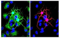
Primary culture of
rat neurons and glial cells.
|
|
Mouse Leydig cell in primary culture.
|
|
|
Tubulin stained by polyclonal antibody (green),
neuron-specific class-III beta tubulin stained with antibody TU-20 (red), DNA (blue).
|
|
Actin fibers stained with
labelled-phalloidin (red), DNA
(blue).
|
|
|
|
|
|
|
|
|
|
|
|
|
|
|
|
|
|
|